NFYA antibody
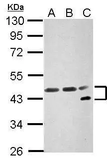
Sample (30 μg of whole cell lysate)
A: NIH-3T3
B: JC
C: BCL-1
10% SDS PAGE
GTX109511 diluted at 1:3000
The HRP-conjugated anti-rabbit IgG antibody (GTX213110-01) was used to detect the primary antibody.
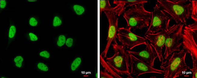
NFYA antibody detects NFYA protein at nucleus by immunofluorescent analysis.Sample: HeLa cells were fixed in 4% paraformaldehyde at RT for 15 min.Green: NFYA stained by NFYA antibody (GTX109511) diluted at 1:500.Red: phalloidin, a cytoskeleton marker, diluted at 1:100.Scale bar= 10 μm.
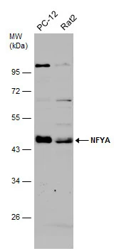
Various whole cell extracts (30 μg) were separated by 10% SDS-PAGE, and the membrane was blotted with NFYA antibody (GTX109511) diluted at 1:3000. The HRP-conjugated anti-rabbit IgG antibody (GTX213110-01) was used to detect the primary antibody.
Various whole cell extracts (30 μg) were separated by 10% SDS-PAGE, and the membrane was blotted with NFYA antibody (GTX109511) diluted at 1:3000. The HRP-conjugated anti-rabbit IgG antibody (GTX213110-01) was used to detect the primary antibody.
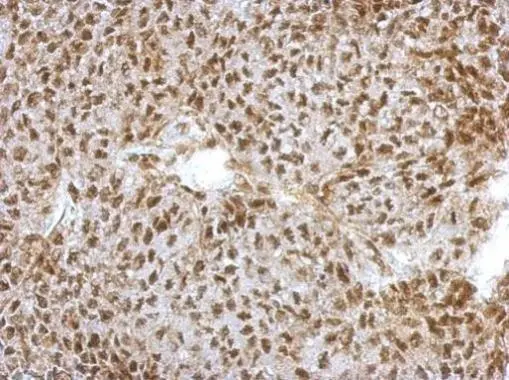
Immunohistochemical analysis of paraffin-embedded D54 xenograft, using NFYA(GTX109511) antibody at 1:500 dilution.
Antigen Retrieval: Trilogy™ (EDTA based, pH 8.0) buffer, 15min
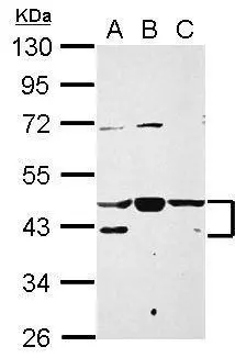
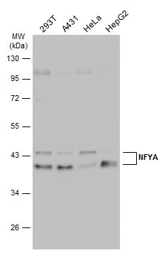
Sample (30 μg of whole cell lysate)
A: A431
B: HeLa
C: HepG2
10% SDS PAGE
GTX109511 diluted at 1:3000
The HRP-conjugated anti-rabbit IgG antibody (GTX213110-01) was used to detect the primary antibody.
-
HostRabbit
-
ClonalityPolyclonal
-
IsotypeIgG
-
ApplicationsWB ICC/IF IHC-P
-
ReactivityHuman, Mouse, Rat