NOL3 antibody

NOL3 antibody detects NOL3 protein at nucleus by immunohistochemical analysis.Sample: Paraffin-embedded mouse brain.NOL3 stained by NOL3 antibody (GTX113889) diluted at 1:500.Antigen Retrieval: Citrate buffer, pH 6.0, 15 min
NOL3 antibody detects NOL3 protein at nucleus by immunohistochemical analysis.Sample: Paraffin-embedded rat brain.NOL3 stained by NOL3 antibody (GTX113889) diluted at 1:500.Antigen Retrieval: Citrate buffer, pH 6.0, 15 min
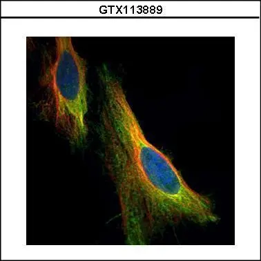
Confocal immunofluorescence analysis (Olympus FV10i) of paraformaldehyde-fixed HeLa, using NOL3(GTX113889) antibody (Green) at 1:500 dilution. Alpha-tubulin filaments were labeled with GTX11304 (Red) at 1:2000.
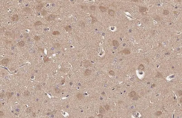
NOL3 antibody detects NOL3 protein at cytoplasm and nucleus by immunohistochemical analysis.Sample: Paraffin-embedded mouse brain.NOL3 stained by NOL3 antibody (GTX113889) diluted at 1:500.
Antigen Retrieval: Citrate buffer, pH 6.0, 15 min
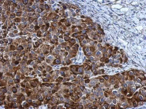
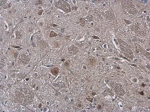
Immunohistochemical analysis of paraffin-embedded Hela xenograft, using NOL3(GTX113889) antibody at 1:500 dilution.
Antigen Retrieval: Trilogy™ (EDTA based, pH 8.0) buffer, 15min
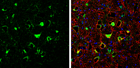
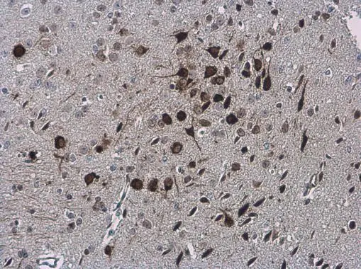
NOL3 antibody detects NOL3 protein by immunohistochemical analysis.Sample: Frozen-sectioned mouse cerebellum.Green: NOL3 stained by NOL3 antibody (GTX113889) diluted at 1:250.Red: NF-H, stained by NF-H antibody [GT114] (GTX634289) diluted at 1:500.Blue: Fluoroshield with DAPI (GTX30920).
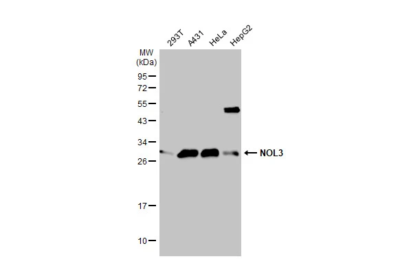
Various whole cell extracts (30 μg) were separated by 12% SDS-PAGE, and the membrane was blotted with NOL3 antibody (GTX113889) diluted at 1:1000. The HRP-conjugated anti-rabbit IgG antibody (GTX213110-01) was used to detect the primary antibody.
NOL3 antibody detects NOL3 protein at cytoplasm and nucleus by immunohistochemical analysis.Sample: Paraffin-embedded rat brain.NOL3 stained by NOL3 antibody (GTX113889) diluted at 1:500.
Antigen Retrieval: Citrate buffer, pH 6.0, 15 min
-
HostRabbit
-
ClonalityPolyclonal
-
IsotypeIgG
-
ApplicationsWB ICC/IF IHC-P IHC-Fr
-
ReactivityHuman, Mouse, Rat