OCC1 antibody
Cat. No. GTX85350
Cat. No. GTX85350
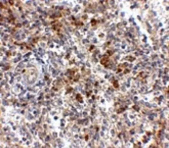
GTX85350 IHC-P Image
IHC-P analysis of human spleen tissue using GTX85350 OCC1 antibody.
Working concentration : 2.5 μg/ml
1 / 3
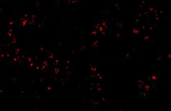
GTX85350 IHC-P Image
IHC-P analysis of human spleen tissue using GTX85350 OCC1 antibody.
Working concentration : 20 μg/ml
2 / 3
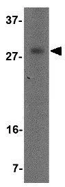
GTX85350 WB Image
WB analysis of human spleen tissue lysate using GTX85350 OCC1 antibody.
Working concentration : 2 μg/ml
3 / 3
-
HostRabbit
-
ClonalityPolyclonal
-
IsotypeIgG
-
ApplicationsWB IHC-P ELISA
-
ReactivityHuman