OCT6 antibody


OCT6 antibody detects OCT6 protein at nucleus by immunohistochemical analysis.Sample: Paraffin-embedded mouse E10.5 embryo.Green: OCT6 stained by OCT6 antibody (GTX134063) diluted at 1:250.Red: beta Tubulin 3/ Tuj1 stained by beta Tubulin 3/ Tuj1 antibody [GT11710] (GTX631836) diluted at 1:500.Blue: Fluoroshield with DAPI (GTX30920).Antigen Retrieval: Citrate buffer, pH 6.0, 15 min
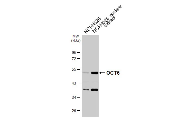
NCI-H526 whole cell and nuclear extracts (30 μg) were separated by 10% SDS-PAGE, and the membrane was blotted with OCT6 antibody (GTX134063) diluted at 1:1000. The HRP-conjugated anti-rabbit IgG antibody (GTX213110-01) was used to detect the primary antibody.

OCT6 antibody detects OCT6 protein at nucleus by immunohistochemical analysis.Sample: Paraffin-embedded mouse E10.5 embryo.Green: OCT6 stained by OCT6 antibody (GTX134063) diluted at 1:500.Red: SOX2, a nucleus marker, stained by SOX2 antibody [GT1352] (GTX627405) diluted at 1:250.Blue: Fluoroshield with DAPI (GTX30920).Antigen Retrieval: Citrate buffer, pH 6.0, 15 min

OCT6 antibody detects OCT6 protein by immunohistochemical analysis.Sample: Frozen-sectioned mouse cerebral cortex.Green: OCT6 stained by OCT6 antibody (GTX134063) diluted at 1:250.Blue: Fluoroshield with DAPI (GTX30920).
-
HostRabbit
-
ClonalityPolyclonal
-
IsotypeIgG
-
ApplicationsWB IHC-P IHC-Fr
-
ReactivityHuman, Mouse, Rat