OSGIN1 antibody
Cat. No. GTX03681
Cat. No. GTX03681
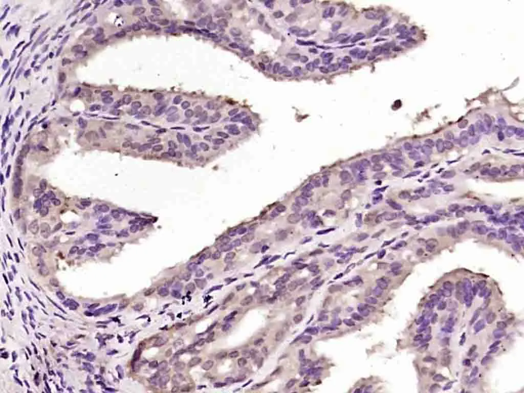
GTX03681 IHC-P Image
IHC-P analysis of rat fallopian tube tissue using GTX03681 OSGIN1 antibody.
Dilution : 1:200
Antigen retrieval : Heat mediated sodium citrate buffer (pH6.0)
1 / 3
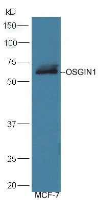
GTX03681 WB Image
WB analysis of MCF-7 cell lysates using GTX03681 OSGIN1 antibody.
Dilution : 1:300
2 / 3
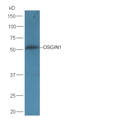
GTX03681 WB Image
WB analysis of mouse liver tissue lysates using GTX03681 OSGIN1 antibody.
Dilution : 1:300
3 / 3
-
HostRabbit
-
ClonalityPolyclonal
-
IsotypeIgG
-
ApplicationsWB IHC-P
-
ReactivityHuman, Mouse, Rat