Optineurin antibody

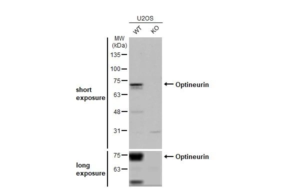
Wild-type (WT) and OPTN knockout (KO) U2OS cell extracts (30 μg) were separated by gradient gel% SDS-PAGE, and the membrane was blotted with Optineurin antibody (GTX132575) diluted at 1:10000. The HRP-conjugated anti-rabbit IgG antibody (GTX213110-01) was used to detect the primary antibody.<br><I>Data provided by YCharOS Inc., an open science company with the mission of characterizing commercially available antibody reagents for all human proteins using knockout technology.</I>
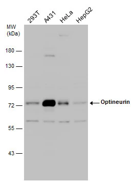
Various whole cell extracts (30 μg) were separated by 7.5% SDS-PAGE, and the membrane was blotted with Optineurin antibody (GTX132575) diluted at 1:2000.
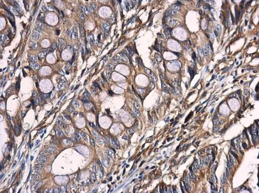
Optineurin antibody detects Optineurin protein at cytoplasm in human colon cancer by immunohistochemical analysis.
Sample: Paraffin-embedded human colon cancer.
Optineurin antibody (GTX132575) diluted at 1:500.
Antigen Retrieval: Citrate buffer, pH 6.0, 15 min
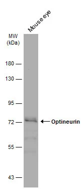
Mouse tissue extract (50 μg) was separated by 7.5% SDS-PAGE, and the membrane was blotted with Optineurin antibody (GTX132575) diluted at 1:500. The HRP-conjugated anti-rabbit IgG antibody (GTX213110-01) was used to detect the primary antibody.
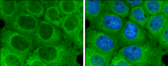
Optineurin antibody detects Optineurin protein at cytoplasm by immunofluorescent analysis.
Sample: A431 cells were fixed in 4% paraformaldehyde at RT for 15 min.
Green: Optineurin protein stained by Optineurin antibody (GTX132575) diluted at 1:500.
-
HostRabbit
-
ClonalityPolyclonal
-
IsotypeIgG
-
ApplicationsWB ICC/IF IHC-P IP
-
ReactivityHuman, Mouse