P2Y1 antibody
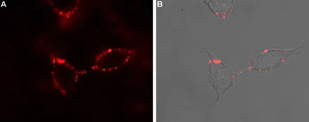
Live cell imaging analysis of intact living PC12 cells using GTX16924 P2Y1 antibody.
Panel A : Primary antibody
Panel B : Merge of panel A with the live view of the cell
Dilution : 1:50
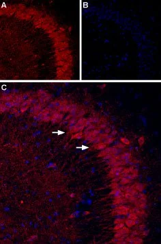
IHC-Fr analysis of mouse hippocampus tissue using GTX16924 P2Y1 antibody.
Panel A : P2RY1 (red) appears in the pyramidal layer (arrows).
Panel B : Nuclei staining using DAPI as the counterstain (blue).
Panel C : Merged images of panels A and B.
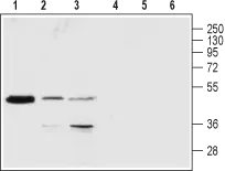
WB analysis of rat brain (lanes 1 and 4), Jurkat (lanes 2 and 5) and K-562 (lanes 3 and 6) lysates using GTX16924 P2Y1 antibody preincubated with or without immunogen peptide.
Dilution : 1:200
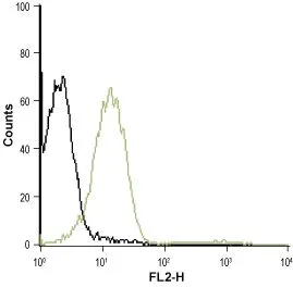
FACS analysis of Jurkat cells using GTX16924 P2Y1 antibody.
Black : Unstained cell
Green : Cell staining with primary antibody
Antibody amount : 5 μg antibody / 0.5-1 x 10⁶ cells
-
HostRabbit
-
ClonalityPolyclonal
-
IsotypeIgG
-
ApplicationsWB ICC/IF IHC-Fr FCM LCI
-
ReactivityHuman, Mouse, Rat