PACSIN1 antibody
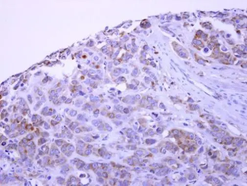
PACSIN1 antibody detects PACSIN1 protein at cytoplasm on MCF7 xenograft by immunohistochemical analysis.
Sample: Paraffin-embedded MCF7 xenograft.
PACSIN1 antibody (GTX108567) dilution: 1:500.
Antigen Retrieval: Trilogy™ (EDTA based, pH 8.0) buffer, 15min
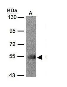
Sample(30 μg of whole cell lysate)
A:MOLT4(GTX27912)
7.5% SDS PAGE
GTX108567 diluted at 1:500
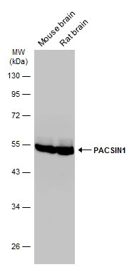
Various tissue extracts (50 μg) were separated by 10% SDS-PAGE, and the membrane was blotted with PACSIN1 antibody (GTX108567) diluted at 1:10000. The HRP-conjugated anti-rabbit IgG antibody (GTX213110-01) was used to detect the primary antibody.
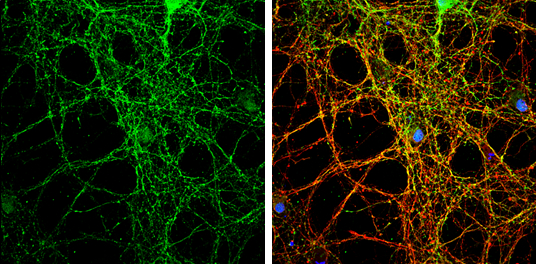
PACSIN1 antibody detects PACSIN1 protein by immunofluorescent analysis.
Sample: DIV14 rat E18 primary cortical neurons were fixed in 4% paraformaldehyde at RT for 15 min.
Green: PACSIN1 protein stained by PACSIN1 antibody (GTX108567) diluted at 1:500.
Red: beta Tubulin 3/ Tuj1, stained by beta Tubulin 3/ Tuj1 antibody [GT1338] (GTX631831) diluted at 1:500.
Blue: Fluoroshield with DAPI (GTX30920).
-
HostRabbit
-
ClonalityPolyclonal
-
IsotypeIgG
-
ApplicationsWB ICC/IF IHC-P
-
ReactivityHuman, Mouse, Rat