PAI2 antibody
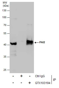
Immunoprecipitation of PAI2 protein from U87-MG whole cell extracts using 5 μg of PAI2 antibody (GTX103194).
Western blot analysis was performed using PAI2 antibody (GTX103194).
EasyBlot anti-Rabbit IgG (GTX221666-01) was used as a secondary reagent.
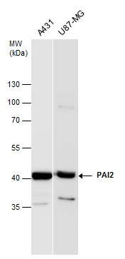
PAI2 antibody detects PAI2 protein by western blot analysis. Various whole cell extracts (30 μg) were separated by 10% SDS-PAGE, and the membrane was blotted with PAI2 antibody (GTX103194) diluted at a dilution of 1:1000. The HRP-conjugated anti-rabbit IgG antibody (GTX213110-01) was used to detect the primary antibody.
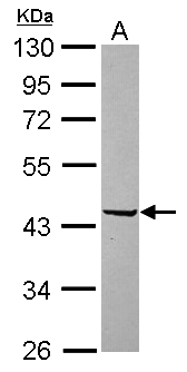
Sample (30 μg of whole cell lysate)
A: U87-MG
10% SDS PAGE
GTX103194 diluted at 1:10000
The HRP-conjugated anti-rabbit IgG antibody (GTX213110-01) was used to detect the primary antibody.
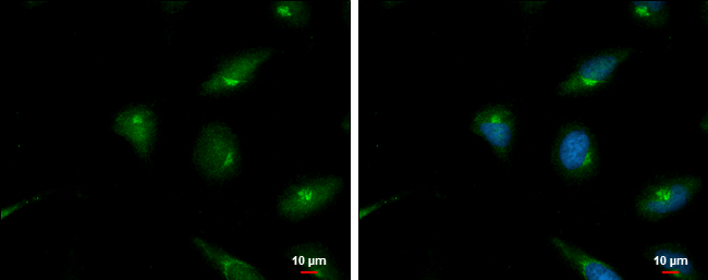
PAI2 antibody detects PAI2 protein at Golgi apparatus by immunofluorescent analysis.
Sample: HeLa cells were fixed in 4% paraformaldehyde at RT for 15 min.
Green: PAI2 protein stained by PAI2 antibody (GTX103194) diluted at 1:500.
Blue: Hoechst 33342 staining.
Scale bar = 10 μm.
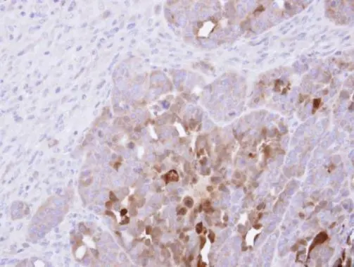
Immunohistochemical analysis of paraffin-embedded NCIN87 xenograft , using PAI-2(GTX103194) antibody at 1:500 dilution.
Antigen Retrieval: Trilogy™ (EDTA based, pH 8.0) buffer, 15min
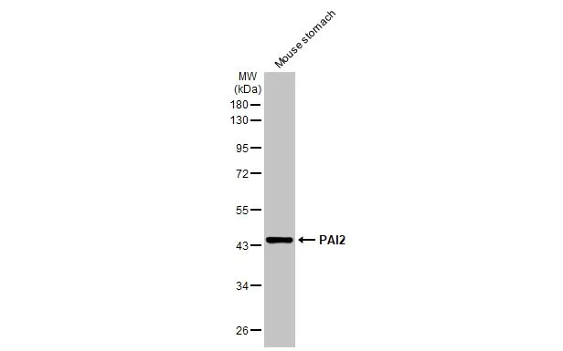
Mouse tissue extract (50 μg) was separated by 10% SDS-PAGE, and the membrane was blotted with PAI2 antibody (GTX103194) diluted at 1:1000. The HRP-conjugated anti-rabbit IgG antibody (GTX213110-01) was used to detect the primary antibody, and the signal was developed with Trident ECL plus-Enhanced.
-
HostRabbit
-
ClonalityPolyclonal
-
IsotypeIgG
-
ApplicationsWB ICC/IF IHC-P IP
-
ReactivityHuman, Mouse