PBK antibody

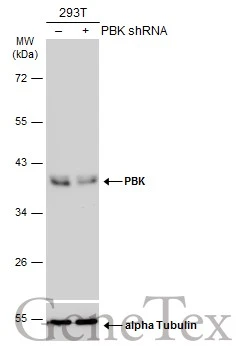
Non-transfected (–) and transfected (+) 293T whole cell extracts (30 μg) were separated by 10% SDS-PAGE, and the membrane was blotted with PBK antibody (GTX131112) diluted at 1:60000. The HRP-conjugated anti-rabbit IgG antibody (GTX213110-01) was used to detect the primary antibody.
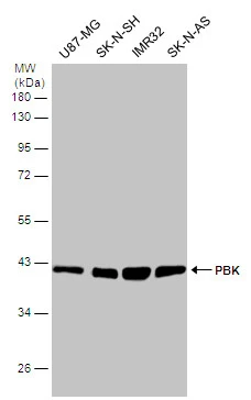
Various whole cell extracts (30 μg) were separated by 10% SDS-PAGE, and the membrane was blotted with PBK antibody (GTX131112) diluted at 1:1000.
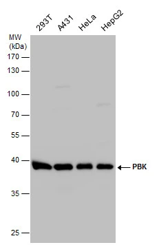
PBK antibody detects PBK protein by western blot analysis. Various whole cell extracts (30 μg) were separated by 10% SDS-PAGE, and the membrane was blotted with PBK antibody (GTX131112) diluted at 1:1000.
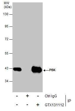
Immunoprecipitation of PBK protein from A431 whole cell extracts using 5 μg of PBK antibody (GTX131112).
Western blot analysis was performed using PBK antibody (GTX131112) diluted at 1:500.
EasyBlot anti-Rabbit IgG (GTX221666-01) was used as a secondary reagent.
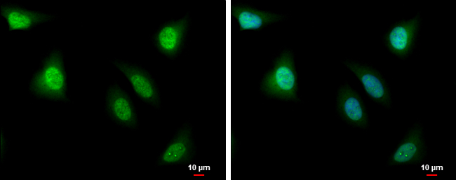
PBK antibody detects PBK protein at nucleus by immunofluorescent analysis.
Sample: HeLa cells were fixed in 4% paraformaldehyde at RT for 15 min.
Green: PBK protein stained by PBK antibody (GTX131112) diluted at 1:500.
Blue: Hoechst 33342 staining.
Scale bar = 10 μm.
-
HostRabbit
-
ClonalityPolyclonal
-
IsotypeIgG
-
ApplicationsWB ICC/IF IP
-
ReactivityHuman