PCNA antibody


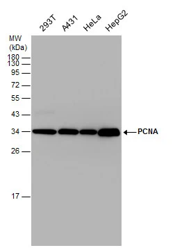
Various whole cell extracts (30 μg) were separated by 12% SDS-PAGE, and the membrane was blotted with PCNA antibody (GTX100539) diluted at 1:2500.
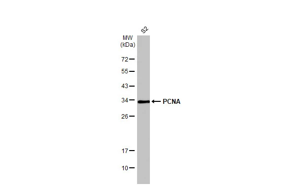
Whole cell extract (30 μg) was separated by 12% SDS-PAGE, and the membrane was blotted with PCNA antibody (GTX100539) diluted at 1:1000. The HRP-conjugated anti-rabbit IgG antibody (GTX213110-01) was used to detect the primary antibody.
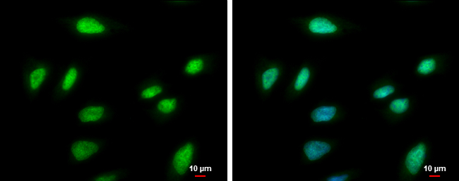
PCNA antibody detects PCNA protein at nucleus by immunofluorescent analysis.
Sample: HeLa cells were fixed in ice-cold MeOH for 5 min.
Green: PCNA protein stained by PCNA antibody (GTX100539) diluted at 1:1000.
Blue: Hoechst 33342 staining.
Scale bar = 10 μm.
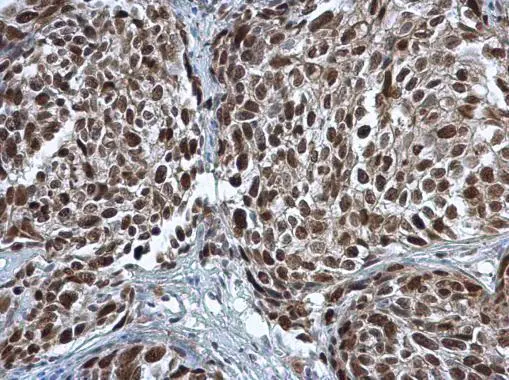
PCNA antibody detects PCNA protein at nucleus in human cervical carcinoma by immunohistochemical analysis.
Sample: Paraffin-embedded human cervical carcinoma.
PCNA antibody (GTX100539) diluted at 1:500.
Antigen Retrieval: Citrate buffer, pH 6.0, 15 min
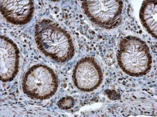
PCNA antibody detects PCNA protein at nucleus in human colon by immunohistochemical analysis.
Sample: Paraffin-embedded human colon .
PCNA antibody (GTX100539) diluted at 1:500.
Antigen Retrieval: Citrate buffer, pH 6.0, 15 min
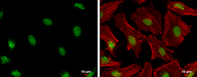
PCNA antibody detects PCNA protein at nucleus by immunofluorescent analysis.
Sample: HeLa cells were fixed in 4% paraformaldehyde at RT for 15 min.
Green: PCNA protein stained by PCNA antibody (GTX100539) diluted at 1:500.
Red: Phalloidin, a cytoskeleton marker, diluted at 1:200.
Scale bar = 10 μm.
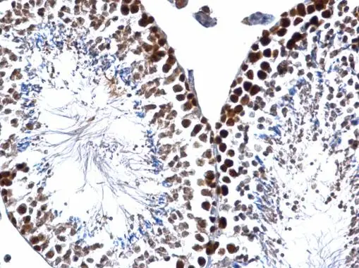
PCNA antibody detects PCNA protein at nucleus on mouse testis by immunohistochemical analysis.
Sample: Paraffin-embedded mouse testis.
PCNA antibody (GTX100539) dilution: 1:500.
Antigen Retrieval: Trilogy™ (EDTA based, pH 8.0) buffer, 15min
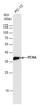
PCNA antibody detects PCNA protein by western blot analysis.
A. 30 μg PC-12 whole cell lysate/extract
12% SDS-PAGE
PCNA antibody (GTX100539) dilution: 1:2500
The HRP-conjugated anti-rabbit IgG antibody (GTX213110-01) was used to detect the primary antibody.
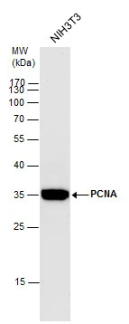
PCNA antibody detects PCNA protein by western blot analysis.
A. 30 μg NIH-3T3 whole cell lysate/extract
12% SDS-PAGE
PCNA antibody (GTX100539) dilution: 1:2500
The HRP-conjugated anti-rabbit IgG antibody (GTX213110-01) was used to detect the primary antibody.
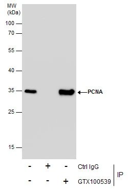
Immunoprecipitation of PCBP2 protein from HeLa whole cell extracts using 5 μg of PCBP2 antibody (GTX100539).
Western blot analysis was performed using PCBP2 antibody (GTX100539).
EasyBlot anti-Rabbit IgG (GTX221666-01) was used as a secondary reagent.
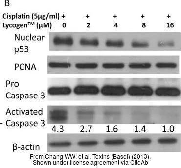
The data was published in the journal Toxins (Basel) in 2013. PMID: 24335753
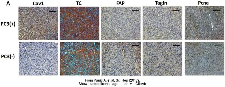
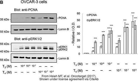
The data was published in the journal Sci Rep in 2017. PMID: 28112237
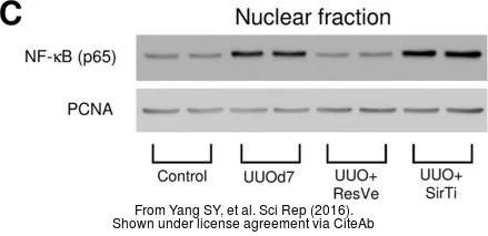
The data was published in the journal Sci Rep in 2016.PMID: 27659793
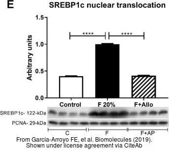
The data was published in the journal Biomolecules in 2019.PMID: 31614639
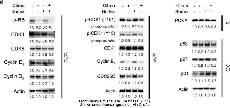
The data was published in the journal Cell Death Dis in 2014.PMID: 25429617
-
HostRabbit
-
ClonalityPolyclonal
-
IsotypeIgG
-
ApplicationsWB ICC/IF IHC-P IP
-
ReactivityHuman, Mouse, Rat, Drosophila, Golden Syrian Hamster