PD-L1 antibody


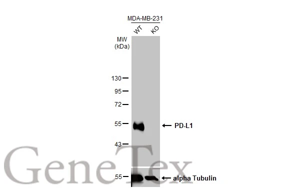
Wild-type (WT) and PD-L1 knockout (KO) MDA-MB-231 cell extracts (30 μg) were separated by 10% SDS-PAGE, and the membrane was blotted with PD-L1 antibody (GTX104763) diluted at 1:4000. The HRP-conjugated anti-rabbit IgG antibody (GTX213110-01) was used to detect the primary antibody.
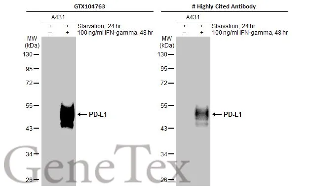
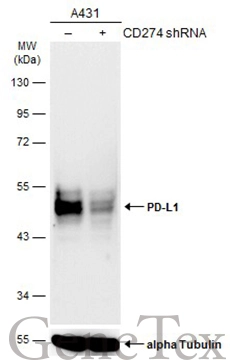
Untreated (–) and treated (+) A431 whole cell extracts (30 μg) were separated by 10% SDS-PAGE, and the membranes were blotted with PD-L1 antibody (GTX104763) diluted at 1:1200 and competitor's antibody diluted at 1:500. The HRP-conjugated anti-rabbit IgG antibody (GTX213110-01) was used to detect the primary antibody.
*The competitor is not affiliated with GeneTex and does not endorse this product.
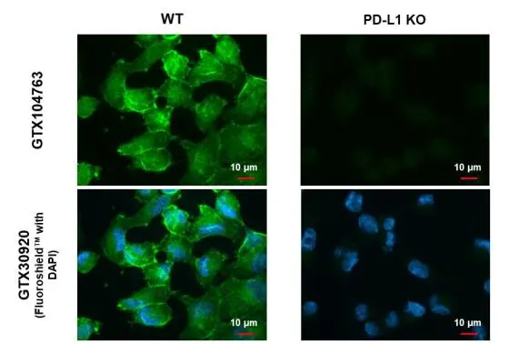
PD-L1 antibody detects PD-L1 protein at cell membrane by immunofluorescent analysis.Sample: MDA-MB-231 cells were fixed in ice-cold MeOH for 5 min.Green: PD-L1 stained by PD-L1 antibody (GTX104763) diluted at 1:500.Blue: Fluoroshield with DAPI (GTX30920).
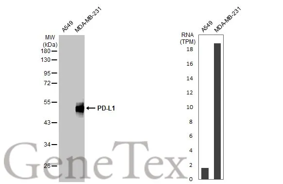
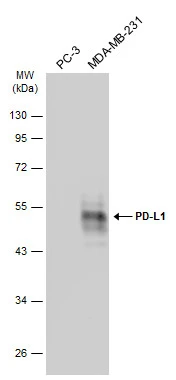
Various whole cell extracts (30 μg) were separated by 10% SDS-PAGE, and the membrane was blotted with PD-L1 antibody (GTX104763) diluted at 1:2000. The HRP-conjugated anti-rabbit IgG antibody (GTX213110-01) was used to detect the primary antibody. Corresponding RNA expression data for the same cell lines are based on Human Protein Atlas program.
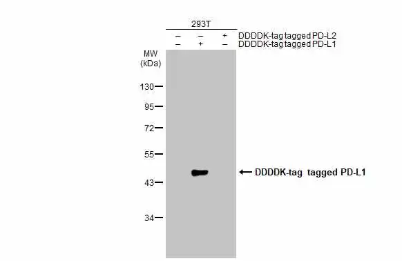
Non-transfected (–) and transfected (+) 293T whole cell extracts (30 μg) were separated by 10% SDS-PAGE, and the membrane was blotted with PD-L1 antibody (GTX104763) diluted at 1:1000. The HRP-conjugated anti-rabbit IgG antibody (GTX213110-01) was used to detect the primary antibody, and the signal was developed with Trident ECL plus-Enhanced.
*The competitor is not affiliated with GeneTex and does not endorse this product.
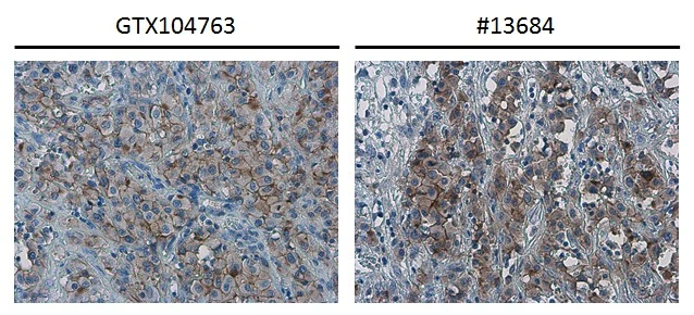
PD-L1 antibody detects PD-L1 protein at cell membrane in human ovarian carcinoma by immunohistochemical analysis. Antibodies: PD-L1 antibody (GTX104763) diluted at 1:1000, and competitor's antibody diluted at 1:50.
Antigen Retrieval: Citrate buffer, pH 6.0, 15 min
*The competitor is not affiliated with GeneTex and does not endorse this product.
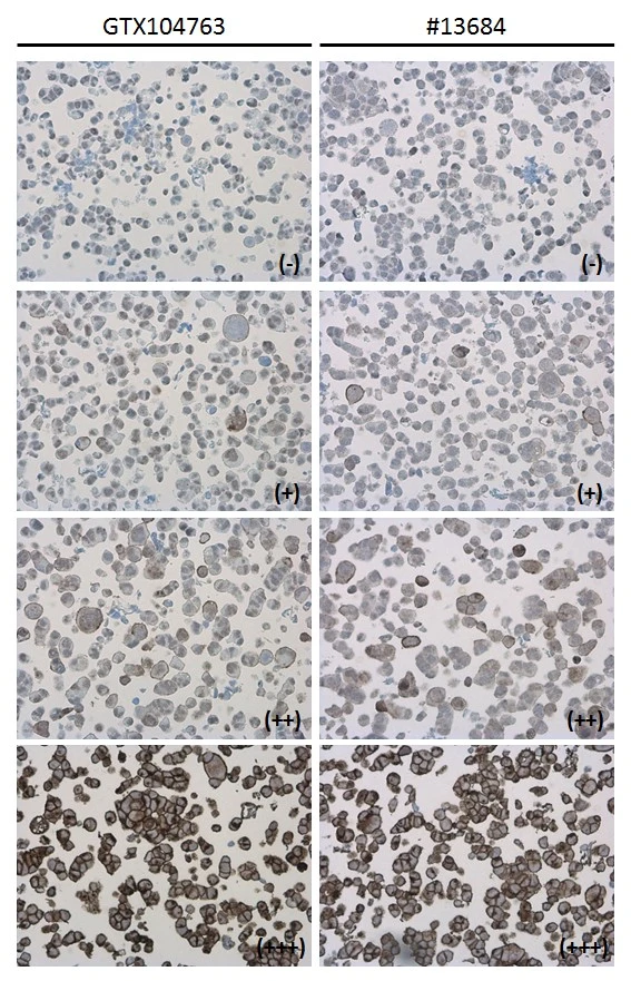
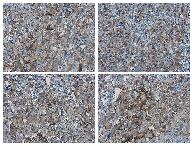
PD-L1 antibody detects PD-L1 protein at cell membrane in PD-L1 protein-expressing cell lines by immunohistochemical analysis. Antibodies: PD-L1 antibody (GTX104763) diluted at 1:1000, and competitor's antibody diluted at 1:50. Samples: Negative (-), low positive (+), intermediate positive (++) and strong positive (+++) cell line cores assessed using Quantitative Digital Pathology.
Antigen Retrieval: Citrate buffer, pH 6.0, 15 min
*The competitor is not affiliated with GeneTex and does not endorse this product.
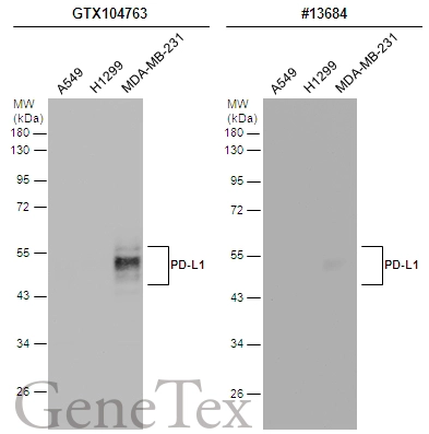
Various whole cell extracts (30 μg) were separated by 12% SDS-PAGE, and the membranes were blotted with PD-L1 antibody (GTX104763) diluted at 1:2000 and competitor's antibody (CST#13684) diluted by 1:500. The HRP-conjugated anti-rabbit IgG antibody (GTX213110-01) was used to detect the primary antibody.
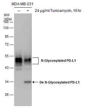
Untreated (–) and treated (+) MDA-MB-231 whole cell extracts (30 μg) were separated by 10% SDS-PAGE, and the membrane was blotted with PD-L1 antibody (GTX104763) diluted at 1:1000.
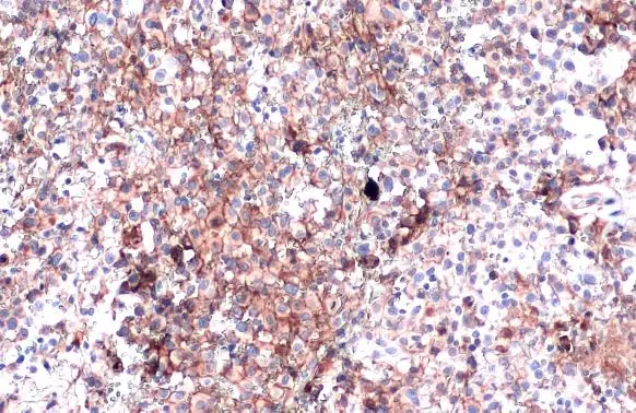
PD-L1 antibody detects PD-L1 proteinat cell membrane in human ovarian carcinoma by immunohistochemical analysis.
Sample: Paraffin-embedded human ovarian carcinoma.
PD-L1 antibody (GTX104763) diluted at 1:1000.
Antigen Retrieval: Citrate buffer, pH 6.0, 15 min
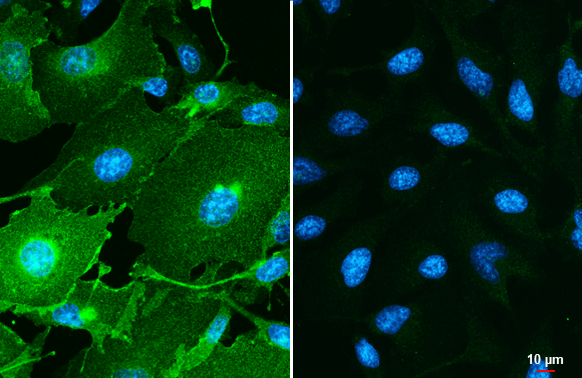
PD-L1 antibody detects PD-L1 protein by immunofluorescent analysis.Sample: MDA-MB-231 (left) and HeLa (right) cells were fixed in ice-cold MeOH for 5 min.Green: PD-L1 stained by PD-L1 antibody (GTX104763) diluted at 1:500.Blue: Hoechst 33342 staining.Scale bar= 10 μm.
PD-L1 antibody detects PD-L1 protein at cell membrane by immunohistochemical analysis.Sample: Paraffin-embedded human ovarian cancer.PD-L1 stained by PD-L1 antibody (GTX104763) diluted at 1:4000.Antigen Retrieval: Citrate buffer, pH 6.0, 15 min

Various whole cell extracts were separated by 10% SDS-PAGE, and the membranes were blotted with PD-L1 antibody (GTX104763) diluted at 1:600 and with DDDDK tag antibody (GTX115043) diluted at 1:3000 to detect DDDDK-tagged PD-L2. The HRP-conjugated anti-rabbit IgG antibody (GTX213110-01) was used to detect the primary antibody.
Non-transfected (–) and transfected (+) A431 whole cell extracts (30 μg) were separated by 10% SDS-PAGE, and the membrane was blotted with PD-L1 antibody (GTX104763) diluted at 1:1000. The HRP-conjugated anti-rabbit IgG antibody (GTX213110-01) was used to detect the primary antibody.
Various whole cell extracts (30 μg) were separated by 10% SDS-PAGE, and the membrane was blotted with PD-L1 antibody (GTX104763) diluted at 1:2000. The HRP-conjugated anti-rabbit IgG antibody (GTX213110-01) was used to detect the primary antibody, and the signal was developed with Trident ECL plus-Enhanced.
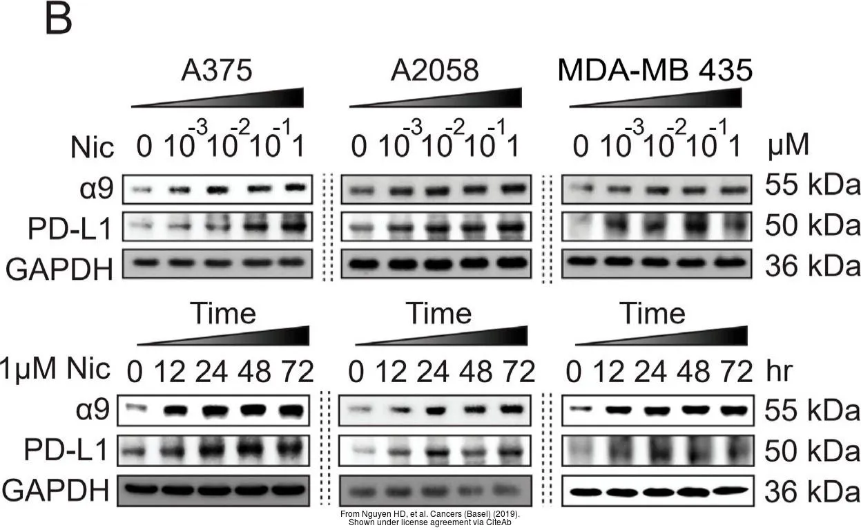
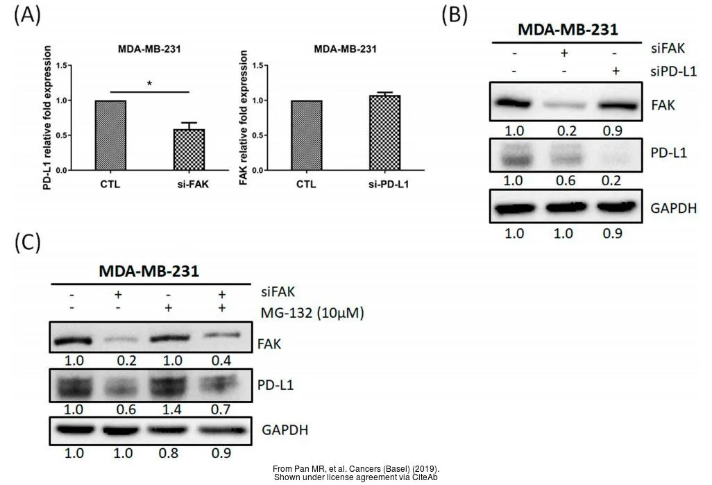
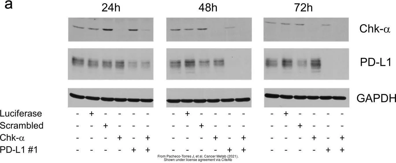
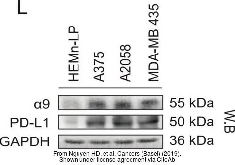
The data was published in the journal Cancers (Basel) in 2019.PMID: 31835799
-
HostRabbit
-
ClonalityPolyclonal
-
IsotypeIgG
-
ApplicationsWB ICC/IF IHC-P IHC-Fr FCM
-
ReactivityHuman