APPLICATION
Application Note
*Optimal dilutions/concentrations should be determined by the researcher.
Application |
Recommended Dilution |
1 μg/mL |
2.5 μg/mL |
Assay dependent |
Not tested in other applications.
Calculated MW
Positive Control
Human Brain Tissue Slide(GTX22203) , THP-1 Cell Lysate(GTX27913)
PROPERTIES
Form
Liquid
Buffer
PBS
Preservative
0.02% Sodium azide
Storage
Store as concentrated solution. Centrifuge briefly prior to opening vial. For short-term storage (1-2 weeks), store at 4ºC. For long-term storage, aliquot and store at -20ºC or below. Avoid multiple freeze-thaw cycles.
Concentration
1 mg/ml (Please refer to the vial label for the specific concentration.)
Antigen Species
Human
Immunogen
PD-1 antibody was raised against a 16 amino acid synthetic peptide from near the carboxy terminus of human PD-1.The immunogen is located within amino acids 210 - 260 of PD-1.
Purification
Purified by antigen-affinity chromatography
Conjugation
Unconjugated
Note
For laboratory use only. Not for any clinical, therapeutic, or diagnostic use in humans or animals. Not for animal or human consumption.
TARGET
Synonyms
programmed cell death 1 , CD279 , PD-1 , PD1 , SLEB2 , hPD-1 , hPD-l , hSLE1
Cellular Localization
Membrane; Single-pass type I membrane protein
Background
This gene encodes a cell surface membrane protein of the immunoglobulin superfamily. This protein is expressed in pro-B-cells and is thought to play a role in their differentiation. In mice, expression of this gene is induced in the thymus when anti-CD3 antibodies are injected and large numbers of thymocytes undergo apoptosis. Mice deficient for this gene bred on a BALB/c background developed dilated cardiomyopathy and died from congestive heart failure. These studies suggest that this gene product may also be important in T cell function and contribute to the prevention of autoimmune diseases. [provided by RefSeq, Jul 2008]
Database
Research Area
DATA IMAGES
|
GTX31756 WB Image
WB analysis of THP-1 cell lysate in the (A) absence and (B) presence of blocking peptide using GTX31756 PD1 antibody. Working concentration : 1 μg/ml
|
|
GTX31756 WB Image
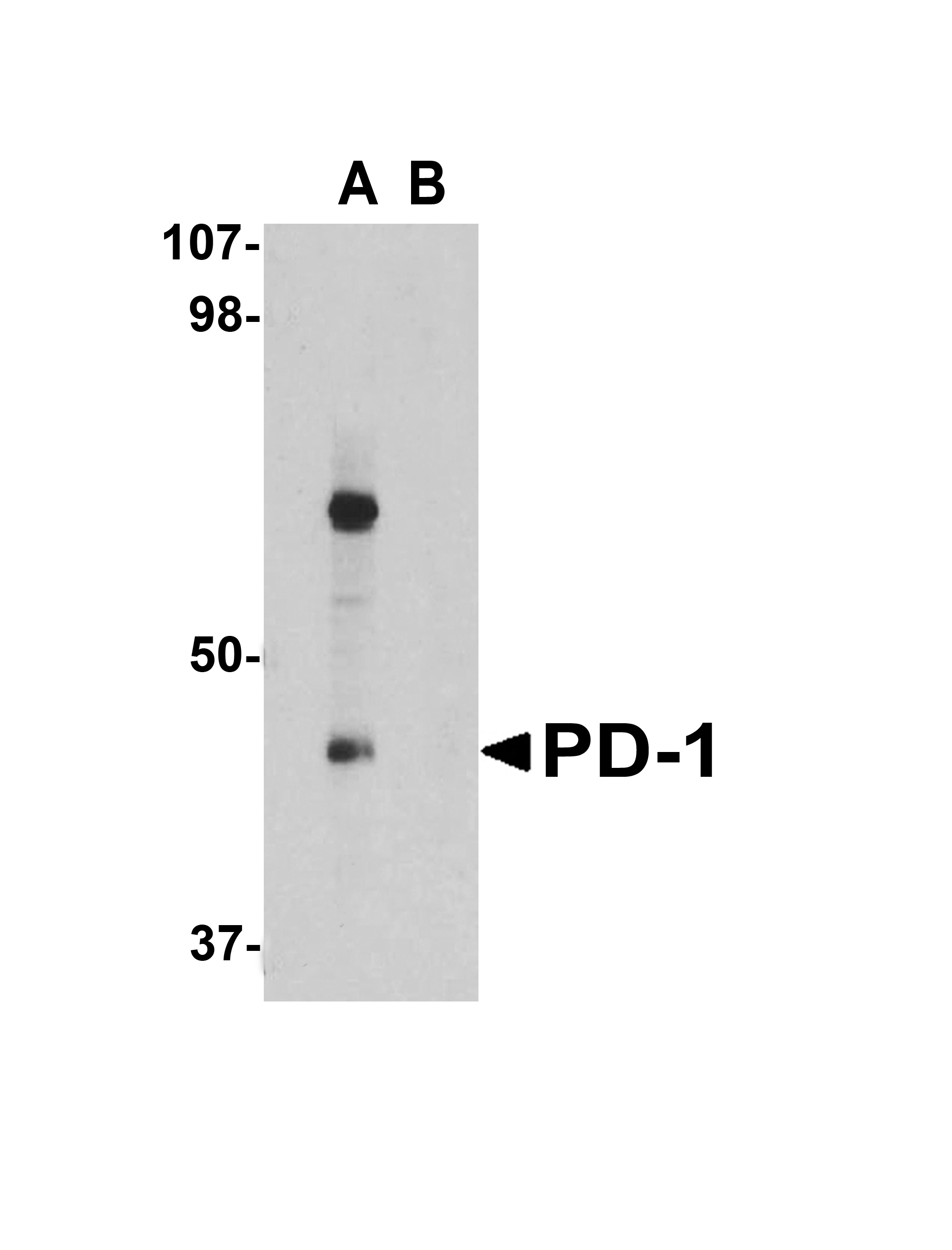
WB analysis of rat thymus cell lysate using GTX31756 PD1 antibody. Dilution : 1 μg/ml
|
|
GTX31756 IHC-P Image
IHC-P analysis of human brain tissue using GTX31756 PD1 antibody. Working concentration : 20 μg/ml
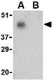
|
|
GTX31756 IHC-P Image
IHC-P analysis of human brain tissue using GTX31756 PD1 antibody. Working concentration : 2.5 μg/ml
|
|
GTX31756 IHC-P Image
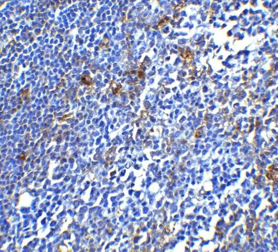
IHC-P analysis of human tonsil tissue using GTX31756 PD1 antibody. Working concentration : 5 μg/ml
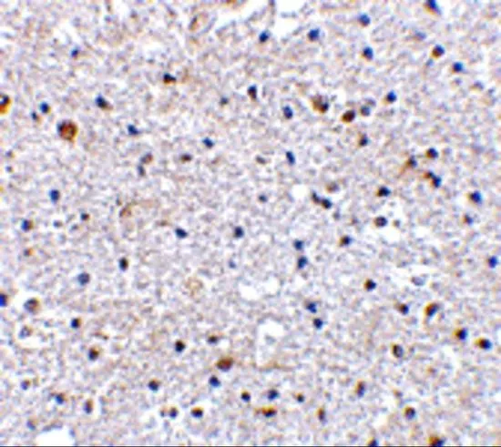
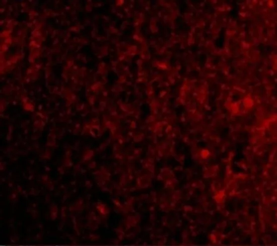
|
REFERENCE
There are currently no references for PD1 antibody (GTX31756). Be the first to share your publications with this product.
REVIEW
There are currently no reviews for PD1 antibody (GTX31756). Be the first to share your experience with this product.