PDCD6 antibody
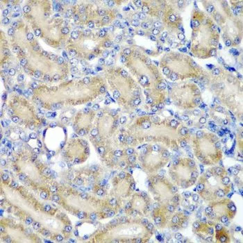
IHC-P analysis of rat kidney tissue using GTX33394 PDCD6 antibody.
Dilution : 1:200
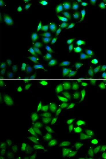
ICC/IF analysis of HeLa cells using GTX33394 PDCD6 antibody.
Blue : DAPI
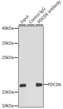
IP analysis of HepG2 cell lysate using GTX33394 PDCD6 antibody.
Antibody amount : 3μg / 200μg lysate
Dilution : 1:1000
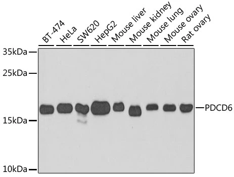
WB analysis of various sample lysates using GTX33394 PDCD6 antibody.
Dilution : 1:1000
Loading : 25μg per lane
-
HostRabbit
-
ClonalityPolyclonal
-
IsotypeIgG
-
ApplicationsWB ICC/IF IHC-P IP
-
ReactivityHuman, Mouse, Rat