APPLICATION
Application Note
*Optimal dilutions/concentrations should be determined by the researcher.
Application |
Recommended Dilution |
1:100-1:1000 |
Not tested in other applications.
Calculated MW
Predict Reactivity
Mouse, Bovine, Rhesus Monkey(>80% identity)
PROPERTIES
Form
Liquid
Buffer
PBS, 20% Glycerol
Preservative
0.01% Thimerosal
Storage
Store as concentrated solution. Centrifuge briefly prior to opening vial. For short-term storage (1-2 weeks), store at 4ºC. For long-term storage, aliquot and store at -20ºC or below. Avoid multiple freeze-thaw cycles.
Concentration
1 mg/ml (Please refer to the vial label for the specific concentration.)
Antigen Species
Human
Immunogen
Recombinant protein encompassing a sequence within the center region of human PDE8A. The exact sequence is proprietary.
Purification
Purified by antigen-affinity chromatography.
Conjugation
Unconjugated
RRID
AB_2885242
Note
For laboratory research use only. Not for any clinical, therapeutic, or diagnostic use in humans or animals. Not for animal or human consumption.
Purchasers shall not, and agree not to enable third parties to, analyze, copy, reverse engineer or otherwise attempt to determine the structure or sequence of the product.
TARGET
Synonyms
phosphodiesterase 8A , HsT19550
Background
Phosphodiesterases (PDEs) regulate the intracellular levels of cAMP and cGMP. These cyclic nucleotides play an important role as second messengers in multiple physiologic processes, including regulation of vascular resistance, cardiac output, visceral motility, immune response, inflammation, neuroplasticity, vision, and reproduction. PDEs comprise a large superfamily of enzymes divided into 10 families. Different PDEs can be distinguished by their structure, tissue expression, localization, substrate specificity, regulation, and sensitivity to PDE inhibitors. Diversity in structure and specificity of function make PDEs promising targets for the pharmacotherapy of diseases modulated by cyclic nucleotide signaling (Hetman et al., MIM 2000). See MIM 171885.[supplied by OMIM]
Database
Research Area
DATA IMAGES
|
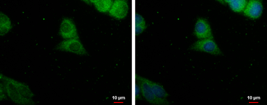
GTX103504 ICC/IF Image
PDE8A antibody detects PDE8A protein at cytoplasm by immunofluorescent analysis. Sample: HepG2 cells were fixed in ice-cold MeOH for 5 min. Green: PDE8A protein stained by PDE8A antibody (GTX103504) diluted at 1:500. Blue: Hoechst 33342 staining.
|
REFERENCE
There are currently no references for PDE8A antibody (GTX103504). Be the first to share your publications with this product.
REVIEW
There are currently no reviews for PDE8A antibody (GTX103504). Be the first to share your experience with this product.