PEG10 antibody
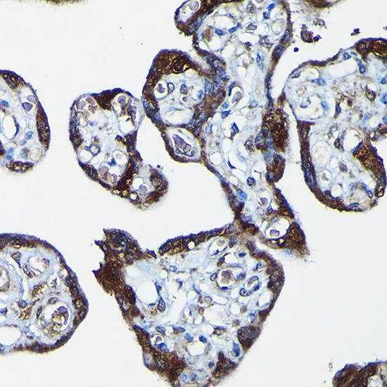
IHC-P analysis of human placenta tissue using GTX64752 PEG10 antibody.
Dilution : 1:100
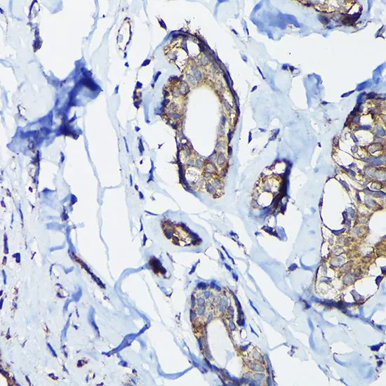
IHC-P analysis of human breast cancer tissue using GTX64752 PEG10 antibody.
Dilution : 1:100
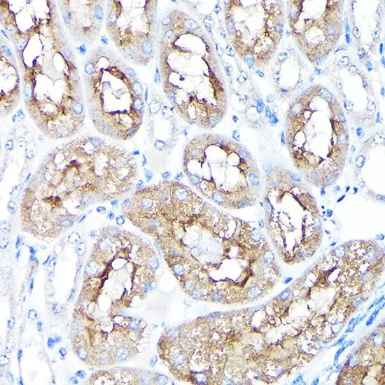
IHC-P analysis of mouse kidney tissue using GTX64752 PEG10 antibody.
Dilution : 1:100
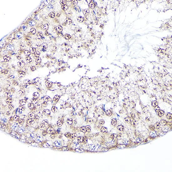
IHC-P analysis of rat testis tissue using GTX64752 PEG10 antibody.
Dilution : 1:100
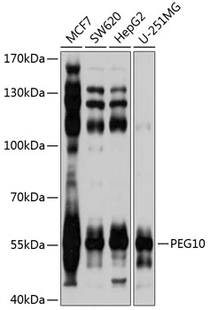
WB analysis of various sample lysates using GTX64752 PEG10 antibody.
Dilution : 1:1000
Loading : 25μg per lane
-
HostRabbit
-
ClonalityPolyclonal
-
IsotypeIgG
-
ApplicationsWB IHC-P
-
ReactivityHuman, Mouse, Rat