PER2 antibody


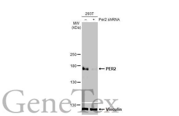
Non-transfected (–) and transfected (+) 293T whole cell extracts (30 μg) were separated by 5% SDS-PAGE, and the membrane was blotted with PER2 antibody (GTX129688) diluted at 1:500. The HRP-conjugated anti-rabbit IgG antibody (GTX213110-01) was used to detect the primary antibody, and the signal was developed with Trident ECL plus-Enhanced.
The observed expression level is based on the publication: PMID: 27834218
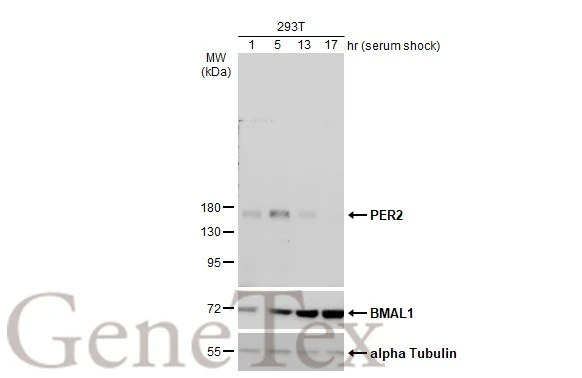
Treated 293T whole cell extracts (30 μg) were separated by 7.5% SDS-PAGE, and the membranes were blotted with PER2 antibody (GTX129688) and BMAL1 antibody [N1N3] (GTX105060) diluted at 1:500. The HRP-conjugated anti-rabbit IgG antibody (GTX213110-01) was used to detect the primary antibody.
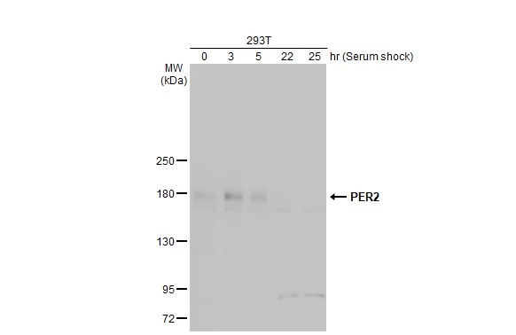
Untreated and treated 293T whole cell extracts (30 μg) were separated by 5% SDS-PAGE, and the membrane was blotted with PER2 antibody (GTX129688) diluted at 1:500. The HRP-conjugated anti-rabbit IgG antibody (GTX213110-01) was used to detect the primary antibody.
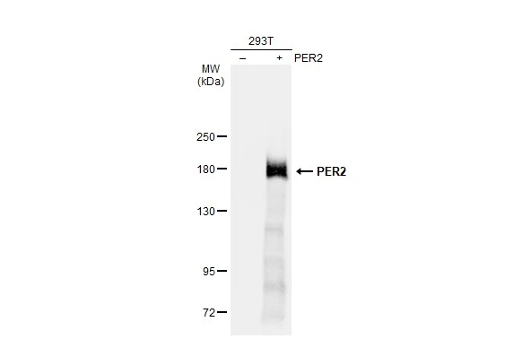
Non-transfected (–) and transfected (+) 293T whole cell extracts (30 μg) were separated by 5% SDS-PAGE, and the membrane was blotted with PER2 antibody (GTX129688) diluted at 1:20000. The HRP-conjugated anti-rabbit IgG antibody (GTX213110-01) was used to detect the primary antibody.
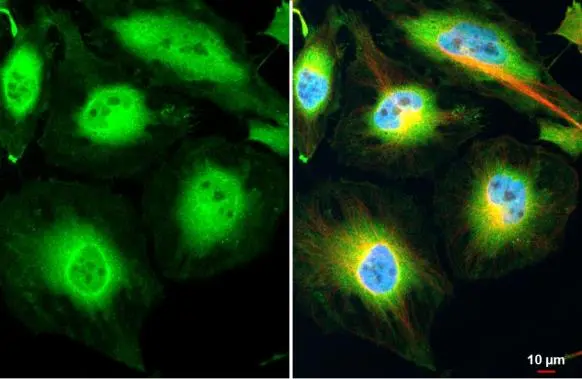
PER2 antibody detects PER2 protein at cytoplasm and nucleus by immunofluorescent analysis.Sample: HeLa cells were fixed in 4% paraformaldehyde at RT for 15 min.Green: PER2 stained by PER2 antibody (GTX129688) diluted at 1:500.Red: alpha Tubulin, a cytoskeleton marker, stained by alpha Tubulin antibody [GT114] (GTX628802) diluted at 1:1000.Blue: Fluoroshield with DAPI (GTX30920).
-
HostRabbit
-
ClonalityPolyclonal
-
IsotypeIgG
-
ApplicationsWB ICC/IF
-
ReactivityHuman