PEX19 antibody

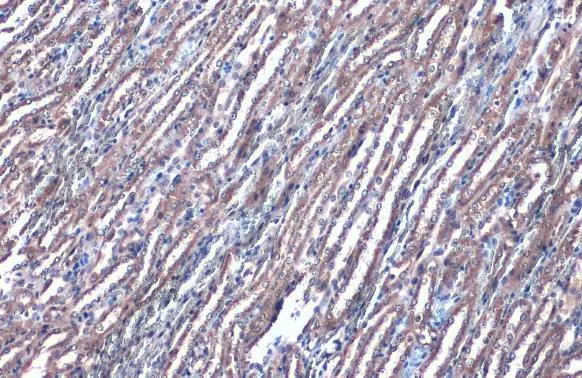
PEX19 antibody detects PEX19 protein at cytoplasm by immunohistochemical analysis.Sample: Paraffin-embedded mouse kidney.PEX19 stained by PEX19 antibody (GTX110721) diluted at 1:1000.Antigen Retrieval: Citrate buffer, pH 6.0, 15 min
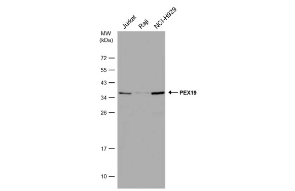
Various whole cell extracts (30 μg) were separated by 12% SDS-PAGE, and the membrane was blotted with PEX19 antibody (GTX110721) diluted at 1:10000. The HRP-conjugated anti-rabbit IgG antibody (GTX213110-01) was used to detect the primary antibody.
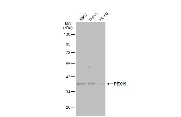
Various whole cell extracts (30 μg) were separated by 10% SDS-PAGE, and the membrane was blotted with PEX19 antibody (GTX110721) diluted at 1:10000. The HRP-conjugated anti-rabbit IgG antibody (GTX213110-01) was used to detect the primary antibody.
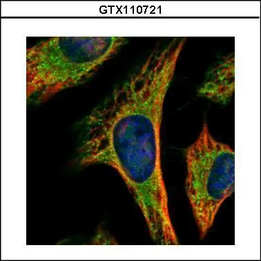
Confocal immunofluorescence analysis (Olympus FV10i) of methanol-fixed HeLa, using PEX19(GTX110721) antibody (Green) at 1:500 dilution. Alpha-tubulin filaments were labeled with GTX11304 (Red) at 1:2000.
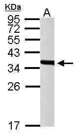
Sample (50 μg of whole cell lysate)
A: mouse liver
12% SDS PAGE
GTX110721 diluted at 1:10000
The HRP-conjugated anti-rabbit IgG antibody (GTX213110-01) was used to detect the primary antibody.
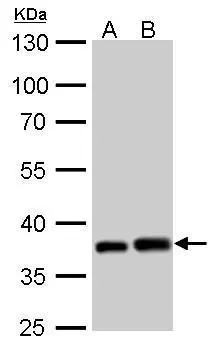
PEX19 antibody detects PEX19 protein by western blot analysis.
A. 30 μg PC-12 whole cell lysate/extract
B. 30 μg Rat-2 whole cell lysate/extract
10% SDS-PAGE
PEX19 antibody (GTX110721) dilution: 1:1000
The HRP-conjugated anti-rabbit IgG antibody (GTX213110-01) was used to detect the primary antibody.
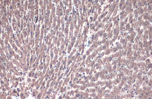
PEX19 antibody detects PEX19 protein at cytoplasm by immunohistochemical analysis.Sample: Paraffin-embedded rat liver.PEX19 stained by PEX19 antibody (GTX110721) diluted at 1:1000.Antigen Retrieval: Citrate buffer, pH 6.0, 15 min
-
HostRabbit
-
ClonalityPolyclonal
-
IsotypeIgG
-
ApplicationsWB ICC/IF IHC-P
-
ReactivityHuman, Mouse, Rat