PGK1 antibody


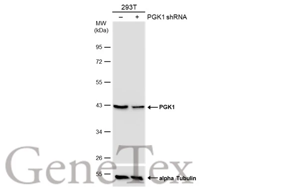
Non-transfected (–) and transfected (+) 293T whole cell extracts (30 μg) were separated by 10% SDS-PAGE, and the membrane was blotted with PGK1 antibody (GTX107614) diluted at 1:1000. The HRP-conjugated anti-rabbit IgG antibody (GTX213110-01) was used to detect the primary antibody.
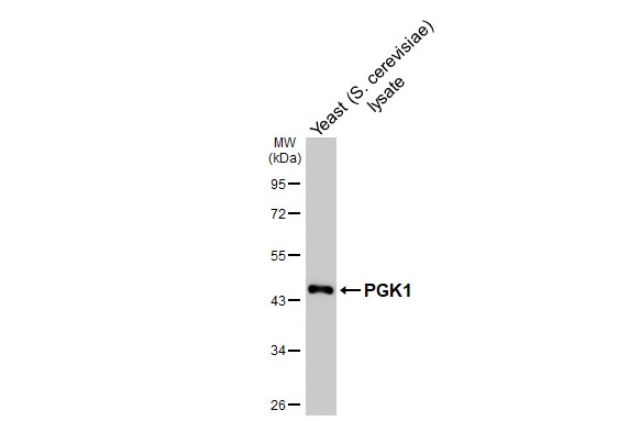
Yeast (S. cerevisiae) lysate was separated by 10% SDS-PAGE, and the membrane was blotted with PGK1 antibody (GTX107614) diluted at 1:1000. The HRP-conjugated anti-rabbit IgG antibody (GTX213110-01) was used to detect the primary antibody.
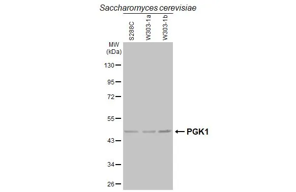
Saccharomyces cerevisiae extracts (30 μg) were separated by 10% SDS-PAGE, and the membrane was blotted with PGK1 antibody (GTX107614) diluted at 1:500. The HRP-conjugated anti-rabbit IgG antibody (GTX213110-01) was used to detect the primary antibody, and the signal was developed with Trident femto Western HRP Substrate.

Mouse tissue extract (50 μg) was separated by 10% SDS-PAGE, and the membrane was blotted with PGK1 antibody (GTX107614) diluted at 1:1000. The HRP-conjugated anti-rabbit IgG antibody (GTX213110-01) was used to detect the primary antibody.

Rat tissue extract (50 μg) was separated by 10% SDS-PAGE, and the membrane was blotted with PGK1 antibody (GTX107614) diluted at 1:1000. The HRP-conjugated anti-rabbit IgG antibody (GTX213110-01) was used to detect the primary antibody, and the signal was developed with Trident femto Western HRP Substrate.
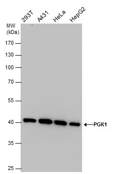
PGK1 antibody detects PGK1 protein by Western blot analysis. Various whole cell extracts (30 μg) were separated by 10% SDS-PAGE, and the membrane was blotted with PGK1 antibody (GTX107614) diluted at a dilution of 1:2000.
PGK1 antibody detects PGK1 protein in 1% O2-treated samples by western blot analysis.
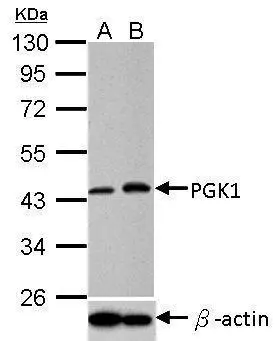
Upper panel: PGK1 antibody (GTX107614)
Lower panel: beta Actin antibody (GTX109639)
A. 30 μg HeLa whole cell lysate/extract (untreated)
B. 30 μg HeLa whole cell lysate/extract (1% O2 treatment for 24hr)
10% SDS PAGE
PGK1 antibody (GTX107614) dilution: 1:1000
beta Actin antibody (GTX109639) dilution: 1:10000
The HRP-conjugated anti-rabbit IgG antibody (GTX213110-01) was used to detect the primary antibody.
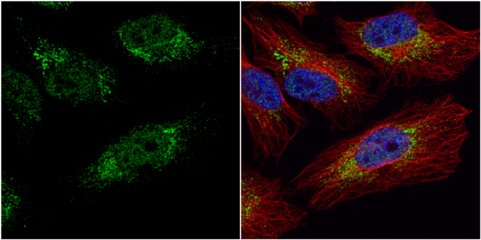
PGK1 antibody detects PGK1 protein at cytoplasm and nucleus by immunofluorescent analysis.
Sample: HeLa cells were fixed in 4% paraformaldehyde at RT for 15 min.
Green: PGK1 protein stained by PGK1 antibody (GTX107614) diluted at 1:20.
Red: alpha Tubulin, a cytoskeleton marker, stained by alpha Tubulin antibody [B-5-1-2] (GTX11304) diluted at 1:10000.
Blue: Hoechst 33342 staining.
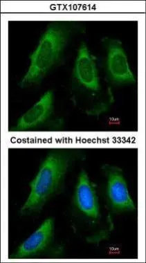
Immunofluorescence analysis of methanol-fixed HeLa, using PGK1(GTX107614) antibody at 1:50 dilution.
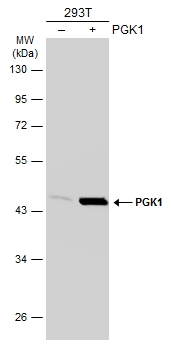
Non-transfected (–) and transfected (+) 293T whole cell extracts (30 μg) were separated by 10% SDS-PAGE, and the membrane was blotted with PGK1 antibody (GTX107614) diluted at 1:3000. The HRP-conjugated anti-rabbit IgG antibody (GTX213110-01) was used to detect the primary antibody.
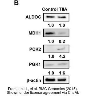
The data was published in the journal BMC Genomics in 2015. PMID: 25652794
-
HostRabbit
-
ClonalityPolyclonal
-
IsotypeIgG
-
ApplicationsWB ICC/IF IP ELISA PLA
-
ReactivityHuman, Mouse, Rat, Yeast