PGM1 antibody
Cat. No. GTX110069
Cat. No. GTX110069
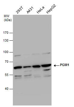
GTX110069 WB Image
PGM1 antibody detects PGM1 protein by western blot analysis. Various whole cell extracts (30 μg) were separated by 7.5% SDS-PAGE, and the membrane was blotted with PGM1 antibody (GTX110069) diluted by 1:500.
1 / 2
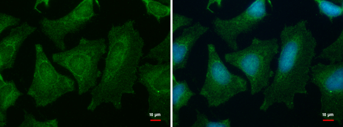
GTX110069 ICC/IF Image
PGM1 antibody detects PGM1 protein at cytoplasm by immunofluorescent analysis.
Sample: HeLa cells were fixed in ice-cold MeOH for 5 min.
Green: PGM1 protein stained by PGM1 antibody (GTX110069) diluted at 1:500.
Blue: Hoechst 33342 staining.
2 / 2
-
HostRabbit
-
ClonalityPolyclonal
-
IsotypeIgG
-
ApplicationsWB ICC/IF
-
ReactivityHuman