PGP9.5 antibody

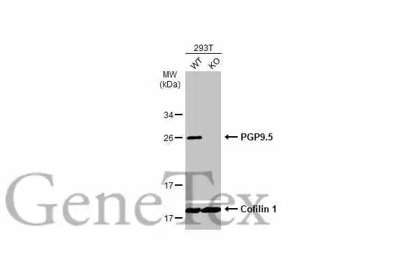
Wild-type (WT) and PGP9.5 knockout (KO) 293T cell extracts (30 μg) were separated by 12% SDS-PAGE, and the membrane was blotted with PGP9.5 antibody (GTX109646) diluted at 1:1000. The HRP-conjugated anti-rabbit IgG antibody (GTX213110-01) was used to detect the primary antibody.
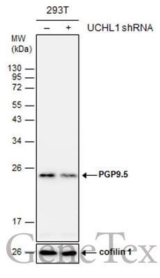
Non-transfected (–) and transfected (+) 293T whole cell extracts (30 μg) were separated by 12% SDS-PAGE, and the membrane was blotted with PGP9.5 antibody (GTX109646) diluted at 1:3000.
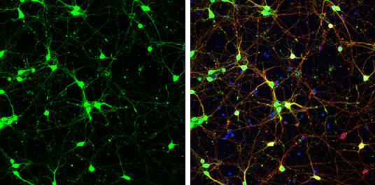
PGP9.5 antibody detects PGP9.5 protein at cytoplasm by immunofluorescent analysis.
Sample: DIV9 rat E18 primary cortical neurons were fixed in 4% paraformaldehyde at RT for 15 min.
Green: PGP9.5 protein stained by PGP9.5 antibody (GTX109646) diluted at 1:500.
Red: beta Tubulin 3/ Tuj1, stained by beta Tubulin 3/ Tuj1 antibody [GT11710] (GTX631836) diluted at 1:500.
Blue: Fluoroshield with DAPI (GTX30920).
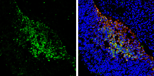
PGP9.5 antibody detects PGP9.5 protein expression by immunohistochemical analysis.
Sample: Frozen sectioned E13.5 Rat brain.
Green: PGP9.5 protein stained by PGP9.5 antibody (GTX109646) diluted at 1:250.
Red: beta Tubulin 3/ TUJ1, a mature neuron marker, stained by beta Tubulin 3/ TUJ1 antibody [GT11710] (GTX631836) diluted at 1:500.
Blue: Fluoroshield with DAPI (GTX30920).
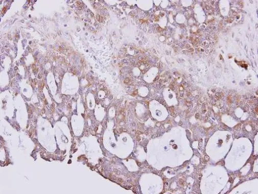
Immunohistochemical analysis of paraffin-embedded SEROUS OVCA xenograft, using PGP9.5(GTX109646) antibody at 1:300 dilution.
Antigen Retrieval: Trilogy™ (EDTA based, pH 8.0) buffer, 15min
PGP9.5 antibody detects UCHL1 protein by Western blot analysis.
A. 50 μg mouse brain lysate/extract
12 % SDS-PAGE
PGP9.5 antibody (GTX109646) dilution: 1:5000
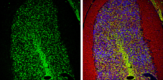
PGP9.5 antibody detects PGP9.5 protein expression by immunohistochemical analysis.
Sample: Frozen-sectioned adult mouse cerebellum.
Green: PGP9.5 protein stained by PGP9.5 antibody (GTX109646) diluted at 1:250.
Red: beta Tubulin 3/ TUJ1, stained by beta Tubulin 3/ TUJ1 antibody [GT11710] (GTX631836) diluted at 1:500.
Blue: Fluoroshield with DAPI (GTX30920).
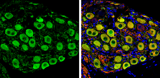
PGP9.5 antibody detects PGP9.5 protein by immunohistochemical analysis.
Samples: Paraffin-embedded rat colon.
Green: PGP9.5 protein stained by PGP9.5 antibody (GTX109646) diluted at 1:250.
Red: beta Tubulin 3/ Tuj1, a marker, stained by beta Tubulin 3/ Tuj1 antibody [GT1338] (GTX631831) diluted at 1:500.
Blue: Fluoroshield with DAPI (GTX30920).
Antigen Retrieval: Citrate buffer, pH 6.0, 15 min
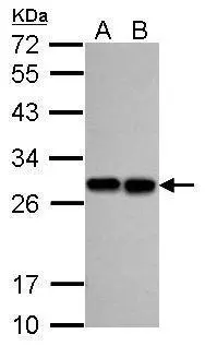
Sample (30 ug of whole cell lysate)
A: A549
B: H1299
12% SDS PAGE
GTX109646 diluted at 1:5000
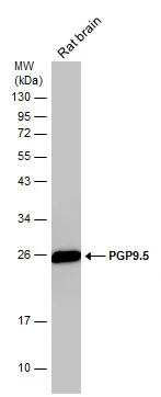
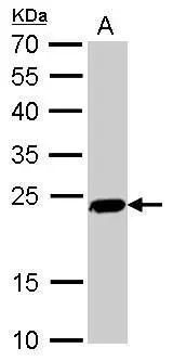
Rat tissue extract (50 μg) was separated by 12% SDS-PAGE, and the membrane was blotted with PGP9.5 antibody (GTX109646) diluted at 1:10000.
-
HostRabbit
-
ClonalityPolyclonal
-
IsotypeIgG
-
ApplicationsWB ICC/IF IHC-P IHC-Fr
-
ReactivityHuman, Mouse, Rat