PKN1 antibody
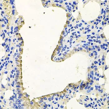
IHC-P analysis of mouse lung tissue using GTX64829 PKN1 antibody.
Dilution : 1:100
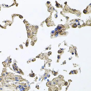
IHC-P analysis of human lung tissue using GTX64829 PKN1 antibody.
Dilution : 1:100
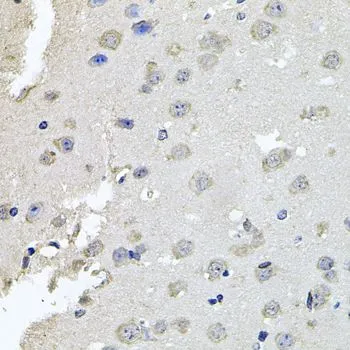
IHC-P analysis of mouse brain tissue using GTX64829 PKN1 antibody.
Dilution : 1:100
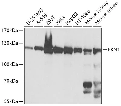
WB analysis of various sample lysates using GTX64829 PKN1 antibody.
Dilution : 1:1000
Loading : 25μg per lane
-
HostRabbit
-
ClonalityPolyclonal
-
IsotypeIgG
-
ApplicationsWB IHC-P
-
ReactivityHuman, Mouse