PMCA1 antibody

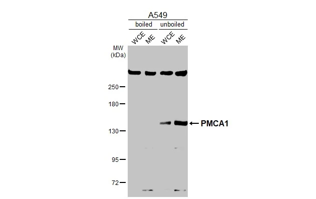
Boiled and unboiled A549 whole cell and membrane extracts (30 μg) were separated by 5% SDS-PAGE, and the membrane was blotted with PMCA1 antibody (GTX130858) diluted at 1:1000. The HRP-conjugated anti-rabbit IgG antibody (GTX213110-01) was used to detect the primary antibody. (WCE: whole cell extract; ME: membrane extract)
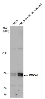
PMCA1 antibody detects PMCA1 protein by western blot analysis. HeLa whole cell extracts and membrane extracts(the extracts were unboiled )(30 μg) were separated by 5% SDS-PAGE, and the membrane was blotted with PMCA1 antibody (GTX130858) at a dilution of 1:1000.
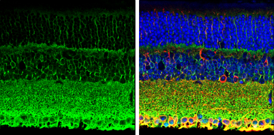
PMCA1 antibody detects PMCA1 protein expression by immunohistochemical analysis.
Sample:Paraffin-Embedded adult mouse retina.
Green: PMCA1 protein stained by PMCA1 antibody (GTX130858) diluted at 1:250.
Red: beta Tubulin 3/ TUJ1, stained by beta Tubulin 3/ TUJ1 antibody [GT11710] (GTX631836) diluted at 1:500.
Blue: Fluoroshield with DAPI (GTX30920).
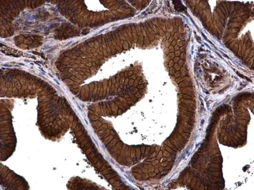
PMCA1 antibody detects PMCA1 protein at cell membrane and cytoplasm in rat prostate by immunohistochemical analysis.
Sample: Paraffin-embedded rat prostate.
PMCA1 antibody (GTX130858) diluted at 1:500.
Antigen Retrieval: Citrate buffer, pH 6.0, 15 min
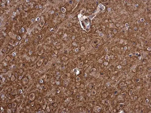
PMCA1 antibody detects PMCA1 protein at cell membrane and cytoplasm in mouse brain by immunohistochemical analysis.
Sample: Paraffin-embedded mouse brain.
PMCA1 antibody (GTX130858) diluted at 1:500.
Antigen Retrieval: Citrate buffer, pH 6.0, 15 min
-
HostRabbit
-
ClonalityPolyclonal
-
IsotypeIgG
-
ApplicationsWB IHC-P IHC-Fr
-
ReactivityHuman, Mouse, Rat