PPAR alpha antibody
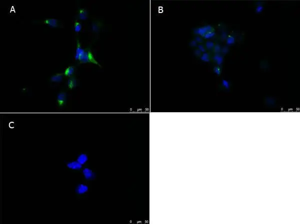
ICC/IF analysis of various MeOH-fixed cells using GTX28934 PPAR alpha antibody.
Panel A : NIH-3T3 cells
Panel B : 293T cells
Panel C : NIH-3T3 cells with secondary antibody only
Green : Primary antibody
Blue : DAPI
Dilution : 10 μg/mL
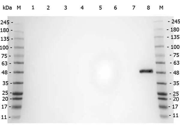
WB analysis of various samples using GTX28934 PPAR alpha antibody.
Lane 1 : 293T whole cell lysate
Lane 2 : HeLa whole cell lysate
Lane 3 : MCF-7 whole cell lysate
Lane 4 : Jurkat whole cell lysate
Lane 5 : A431 whole cell lysate
Lane 6 : LNCaP whole cell lysate
Lane 7 : A-172 whole cell lysate
Lane 8 : NIH-3T3 whole cell lysate
Loading : 35 μg
Dilution : 1 μg/mL
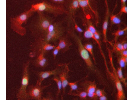
ICC/IF analysis of PFA-fixed HepG2 cells using GTX28934 PPAR alpha antibody.
Green : Primary antibody
Red : Plasma membrane
Blue : DAPI
Dilution : 1 μg/mL
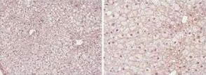
IHC-P analysis of mouse liver tissue using GTX28934 PPAR alpha antibody.
Antigen retrieval : 0.05% protease in PBS
Dilution : 1:50
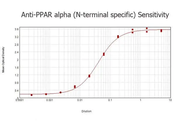
ELISA analysis of BSA-conjugated immunizing peptide using serially diluted GTX28934 PPAR alpha antibody.
Coating : 0.1 μg
WB analysis of NIH-3T3 whole cell lysate using GTX28934 PPAR alpha antibody.
Lane 1 : 1:500
Lane 2 : 1:1000
Loading : 20 μg
WB analysis of NIH-3T3 whole cell lysate using GTX28934 PPAR alpha antibody.
Loading : 10 μg
Dilution : 1:1000
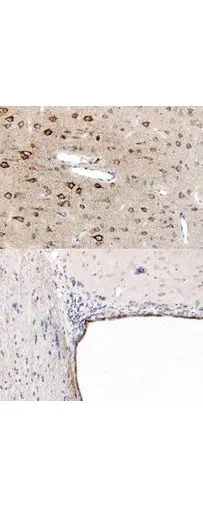
Immunohistochemistry using GeneTex's anti-PPAR antibody, showing staining of PPAR alpha in rat brain sections, highlighting cytoplasmic staining in ependymal cells and neurons in frontal cortex. Bottom image shows subventricular zone (svz) of lateral ventrical (exit point of progenitor olfactory neurones); top image shows frontal cortex in the same section. Cytoplasmic staining is also observed in the corpus callosum (bottom image) and in dendritic fields of the cortex. Formalin/PFA-fixed paraffin-embedded sections of rat brain tissue were incubated with the primary antibody at 1:200 for 1 hour. Antigen retrieval was performed by heat induction in citrate buffer pH 6.0.

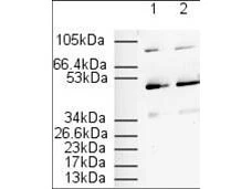
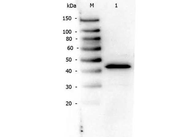
Affinity Purified anti-PPAR alpha (N -terminal specific) (Rabbit) is shown to detect a 52 kDa band corresponding to PPAR alpha present in a 3T3 whole cell lysate. Approximately 20 μg of lysate was loaded per lane for SDS-PAGE. Detection occurred after using a 1:500 (lane 1) or 1:1000 (lane 2) dilution of antibody followed by 1:2000 dilution of HRP Goat-a-Rabbit IgG for visualization.
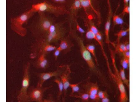
Immunofluorescence Microscopy of Rabbit anti-PPAR alpha antibody (GTX28934). Tissue: HepG2 cells. Fixation: 4% formaldehyde fixed (10 min). Antigen retrieval: not required. Primary antibody: PPAR alpha antibody at 1 μg/mL overnight at 4ºC. Secondary antibody: Alexa FluorR 488 goat anti-rabbit IgG (H+L) (green) used at a 1:1000, Alexa FluorR 594 WGA was used to label plasma membranes (red) at a 1:200 dilution for 1h for 45 min at RT. Localization: PPAR alpha is nuclear and occasionally cytoplasmic. Staining: PPAR alpha as green fluorescent signal with DAPI (blue) nuclear counterstain.
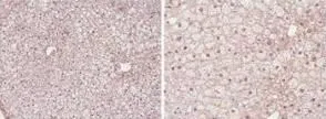
Immunohistochemistry showing GeneTex's PPAR alpha antibody staining of PPAR alpha protein in mouse liver tissue section (Formalin/PFA-fixed paraffin-embedded sections). Tissue underwent formaldehyde fixation before enzymatic antigen retrieval with 0.05% protease in PBS for 5 minutes. Sample was then blocked with 5% serum for 20 minutes at 20ºC. The primary antibody was diluted 1:50 and incubated with sample in Tris plus 5% normal goat serum for 1 hour at 20ºC. A biotinylated goat polyclonal to rabbit IgG was used at dilution at 1:500 as secondary antibody. Images show nuclear staining in hepatocytes (perfusion-fixed mouse, 10 and 40x microscope magnification).
-
HostRabbit
-
ClonalityPolyclonal
-
IsotypeIgG
-
ApplicationsWB ICC/IF IHC-P FCM ELISA ChIP assay
-
ReactivityHuman, Mouse, Rat