PRDM4 antibody

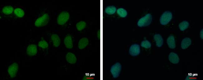
PRDM4 antibody detects PRDM4 protein at nucleus by immunofluorescent analysis.
Sample: NT2D1 cells were fixed in 4% paraformaldehyde at RT for 15 min.
Green: PRDM4 protein stained by PRDM4 antibody (GTX115579) diluted at 1:500.
Blue: Hoechst 33342 staining.
Scale bar = 10 μm.
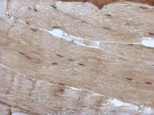
Immunohistochemical analysis of paraffin-embedded mouse muscle, using PRDM4(GTX115579) antibody at 1:500 dilution.
Antigen Retrieval: Trilogy™ (EDTA based, pH 8.0) buffer, 15min
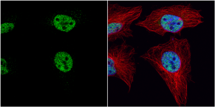
PRDM4 antibody detects PRDM4 protein at nucleus by immunofluorescent analysis.
Sample: HeLa cells were fixed in 4% paraformaldehyde at RT for 15 min.
Green: PRDM4 protein stained by PRDM4 antibody (GTX115579) diluted at 1:1000.
Red: alpha Tubulin, a cytoskeleton marker, stained by alpha Tubulin antibody [GT114] (GTX628802) diluted at 1:1000.
Blue: Hoechst 33342 staining.
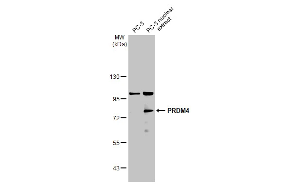
PC-3 whole cell and nuclear extracts (30 μg) were separated by 7.5% SDS-PAGE, and the membrane was blotted with PRDM4 antibody (GTX115579) diluted at 1:50000. The HRP-conjugated anti-rabbit IgG antibody (GTX213110-01) was used to detect the primary antibody.
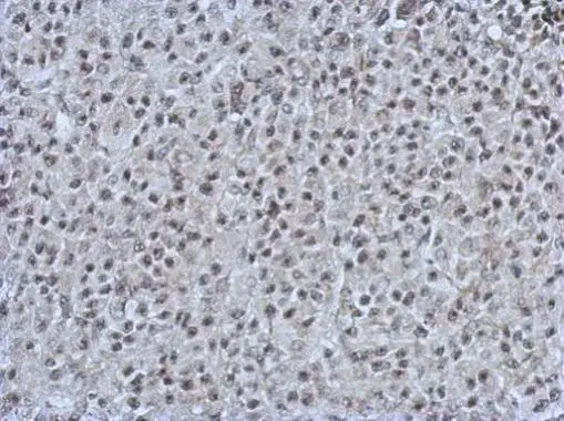
Immunohistochemical analysis of paraffin-embedded HBL435 xenograft, using PRDM4(GTX115579) antibody at 1:500 dilution.
Antigen Retrieval: Trilogy™ (EDTA based, pH 8.0) buffer, 15min
-
HostRabbit
-
ClonalityPolyclonal
-
IsotypeIgG
-
ApplicationsWB ICC/IF IHC-P
-
ReactivityHuman, Mouse