PSMD2 antibody
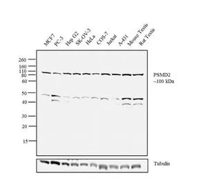
WB analysis of MCF7 (Lane 1), PC-3 (Lane 2), Hep G2 (Lane 3), SK-OV-3 (Lane 4), HeLa (Lane 5), COS-7 (Lane 6), Jurkat (Lane 7), A-431 (Lane 8), tissue extracts of mouse testis (Lane 9) and rat testis (Lane 10) using GTX79499 PSMD2 antibody.
Dilution : 1:1000
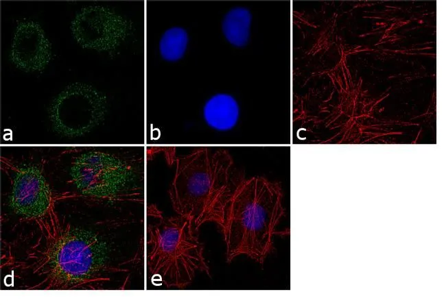
ICC/IF analysis of HepG2 cells using GTX79499 PSMD2 antibody. Panel e is a no primary antibody control.
Green : Primary antibody
Blue : Nuclei
Red : Actin
Fixation : 4% paraformaldehyde
Permeabilization : 0.1% Triton X-100 for 10 minute
Dilution : 1:250 dilution in 0.1% BSA and incubated for 3 hours at room temperature
-
HostRabbit
-
ClonalityPolyclonal
-
IsotypeIgG
-
ApplicationsWB ICC/IF
-
ReactivityHuman, Mouse, Rat, Dog, Hamster, Monkey, Primate