PTEN antibody


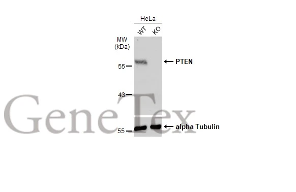
Wild-type (WT) and PTEN knockout (KO) HeLa cell extracts (30 μg) were separated by 7.5% SDS-PAGE, and the membrane was blotted with PTEN antibody (GTX101025) diluted at 1:500. The HRP-conjugated anti-rabbit IgG antibody (GTX213110-01) was used to detect the primary antibody.
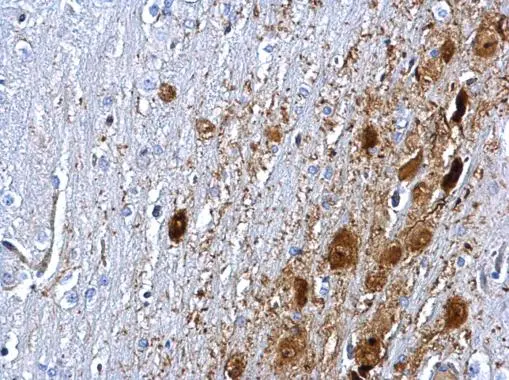
PTEN antibody detects PTEN protein at cytosol and nucleus on rat middle brain by immunohistochemical analysis.
Sample: Paraffin-embedded rat middle brain.
PTEN antibody (GTX101025) dilution: 1:500.
Antigen Retrieval: Trilogy™ (EDTA based, pH 8.0) buffer, 15min
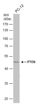
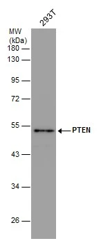
Whole cell extract (30 μg) was separated by 10% SDS-PAGE, and the membrane was blotted with PTEN antibody (GTX101025) diluted at 1:500. The HRP-conjugated anti-rabbit IgG antibody (GTX213110-01) was used to detect the primary antibody.
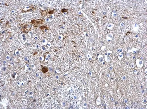
PTEN antibody detects PTEN protein at cytosol and nucleus on mouse middle brain by immunohistochemical analysis.
Sample: Paraffin-embedded mouse middle brain.
PTEN antibody (GTX101025) dilution: 1:500.
Antigen Retrieval: Trilogy™ (EDTA based, pH 8.0) buffer, 15min
Whole cell extract (30 μg) was separated by 10% SDS-PAGE, and the membrane was blotted with PTEN antibody (GTX101025) diluted at 1:500. The HRP-conjugated anti-rabbit IgG antibody (GTX213110-01) was used to detect the primary antibody.
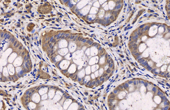
PTEN antibody detects PTEN protein at cytoplasm by immunohistochemical analysis (Autostainer Formulated).Sample: Paraffin-embedded human normal colon tissue.PTEN stained by PTEN antibody (GTX101025) diluted at 1:500.Antigen Retrieval: Citrate buffer, 20 min

PTEN antibody detects PTEN protein at cytoplasm in human ovarian cancer by immunohistochemical analysis.
Sample: Paraffin-embedded human ovarian cancer.
PTEN antibody (GTX101025) diluted at 1:500.
Antigen Retrieval: Citrate buffer, pH 6.0, 15 min
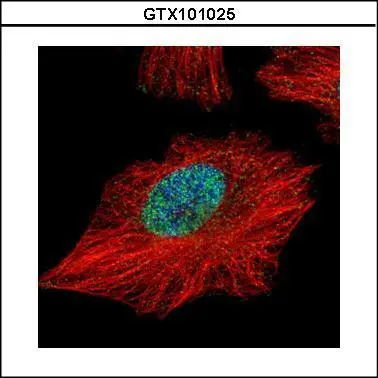
Confocal immunofluorescence analysis (Olympus FV10i) of paraformaldehyde-fixed HeLa, using PTEN(GTX101025) antibody (Green) at 1:500 dilution. Alpha-tubulin filaments were labeled with GTX11304 (Red) at 1:2000.
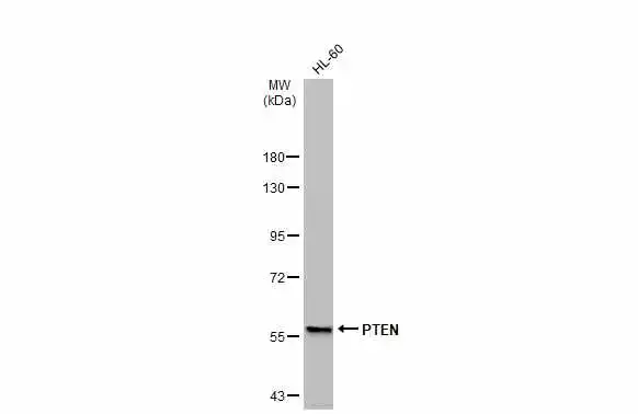
Whole cell extract (30 μg) was separated by 7.5% SDS-PAGE, and the membrane was blotted with PTEN antibody (GTX101025) diluted at 1:500. The HRP-conjugated anti-rabbit IgG antibody (GTX213110-01) was used to detect the primary antibody.
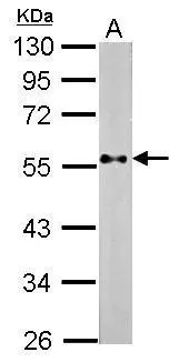
Sample (30 μg of whole cell lysate)
A: NIH-3T3
10% SDS PAGE
GTX101025 diluted at 1:500
The HRP-conjugated anti-rabbit IgG antibody (GTX213110-01) was used to detect the primary antibody.
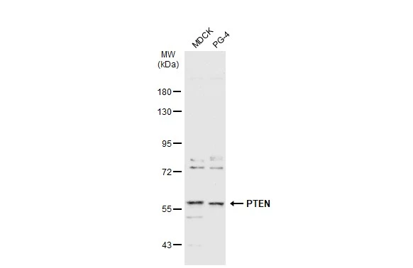
Various whole cell extracts (30 μg) were separated by 7.5% SDS-PAGE, and the membrane was blotted with PTEN antibody (GTX101025) diluted at 1:1000. The HRP-conjugated anti-rabbit IgG antibody (GTX213110-01) was used to detect the primary antibody.
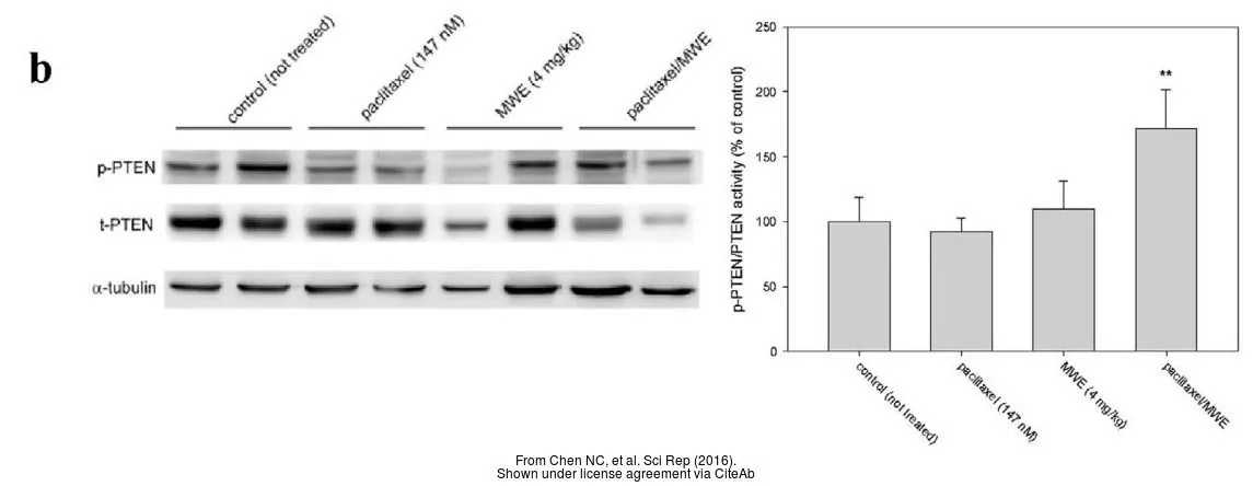
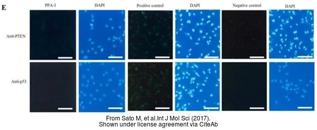
The data was published in the journal Int J Mol Sci in 2017. PMID: 29207527
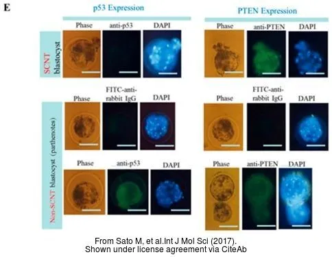
The data was published in the journal Int J Mol Sci in 2017. PMID: 29207527
-
HostRabbit
-
ClonalityPolyclonal
-
IsotypeIgG
-
ApplicationsWB ICC/IF IHC-P
-
ReactivityHuman, Mouse, Rat, Cat, Dog, Pig