GeneTex
  • Country / Location Selection

United States (US)

Pentraxin 3 antibody

Anti-Pentraxin 3 antibody used in Western Blot (WB). GTX135596
Anti-Pentraxin 3 antibody used in Immunocytochemistry/ Immunofluorescence (ICC/IF). GTX135596

Cat. No. GTX135596

Host

Rabbit

Clonality

Polyclonal

Isotype

IgG

Application

WB, ICC/IF

Reactivity

Human
Package
100 μl ($399),
25 μl ($169)

APPLICATION

Application Note

*Optimal dilutions/concentrations should be determined by the researcher.
Application Recommended Dilution
WB 1:500-1:3000
ICC/IF Assay dependent
Not tested in other applications.

Calculated MW

42 kDa. ( Note )

Predict Reactivity

Mouse, Pig(>80% identity)

PROPERTIES

Form

Liquid

Buffer

PBS, 20% Glycerol

Preservative

0.025% ProClin 300

Storage

Store as concentrated solution. Centrifuge briefly prior to opening vial. For short-term storage (1-2 weeks), store at 4ºC. For long-term storage, aliquot and store at -20ºC or below. Avoid multiple freeze-thaw cycles.

Concentration

0.5 mg/ml (Please refer to the vial label for the specific concentration.)

Antigen Species

Human

Immunogen

Recombinant protein encompassing a sequence within the center region of human Pentraxin 3. The exact sequence is proprietary.

Purification

Purified by antigen-affinity chromatography.

Conjugation

Unconjugated

RRID

AB_2909858

Note

For laboratory research use only. Not for any clinical, therapeutic, or diagnostic use in humans or animals. Not for animal or human consumption.

Purchasers shall not, and agree not to enable third parties to, analyze, copy, reverse engineer or otherwise attempt to determine the structure or sequence of the product.

TARGET

Synonyms

pentraxin 3 , TNFAIP5 , TSG-14

Cellular Localization

Secreted

Background

This gene encodes a member of the pentraxin protein family. The expression of this protein is induced by inflammatory cytokines in response to inflammatory stimuli in several mesenchymal and epithelial cell types, particularly endothelial cells and mononuclear phagocytes. The protein promotes fibrocyte differentiation and is involved in regulating inflammation and complement activation. It also plays a role in angiogenesis and tissue remodeling. The protein serves as a biomarker for several inflammatory conditions. [provided by RefSeq, Jun 2016]

Database

Research Area

DATA IMAGES

Anti-Pentraxin 3 antibody used in Western Blot (WB). GTX135596

GTX135596 WB Image

Whole cell extract (30 μg) was separated by 10% SDS-PAGE, and the membrane was blotted with Pentraxin 3 antibody (GTX135596) diluted at 1:3000. The HRP-conjugated anti-rabbit IgG antibody (GTX213110-01) was used to detect the primary antibody.

Anti-Pentraxin 3 antibody used in Immunocytochemistry/ Immunofluorescence (ICC/IF). GTX135596

GTX135596 ICC/IF Image

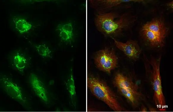
Pentraxin 3 antibody detects Pentraxin 3 protein at mitochondria by immunofluorescent analysis.
Sample: HUVEC cells were fixed in 4% paraformaldehyde at RT for 15 min.
Green: Pentraxin 3 stained by Pentraxin 3 antibody (GTX135596) diluted at 1:500.
Red: alpha Tubulin, a cytoskeleton marker, stained by alpha Tubulin antibody [GT114] (GTX628802) diluted at 1:1000.
Blue: Fluoroshield with DAPI (GTX30920).
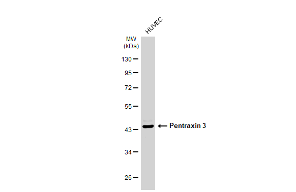
REFERENCE

There are currently no references for Pentraxin 3 antibody (GTX135596). Be the first to share your publications with this product.

REVIEW

There are currently no reviews for Pentraxin 3 antibody (GTX135596). Be the first to share your experience with this product.
Package List Price ($)
$ 399
$ 169