GeneTex
  • Country / Location Selection

United States (US)

Perilipin 1 antibody

Anti-Perilipin 1 antibody used in Western Blot (WB). GTX130139
Anti-Perilipin 1 antibody used in IHC (Paraffin sections) (IHC-P). GTX130139

Cat. No. GTX130139

Host

Rabbit

Clonality

Polyclonal

Isotype

IgG

Application

WB, IHC-P

Reactivity

Mouse
Package
100 μl ($399),
25 μl ($169)

APPLICATION

Application Note

*Optimal dilutions/concentrations should be determined by the researcher.
Application Recommended Dilution
WB 1:500-1:10000
IHC-P Assay dependent
Not tested in other applications.

Calculated MW

56 kDa. ( Note )

Predict Reactivity

Human, Rat, Sheep, Bovine, Pig(>80% identity)

PROPERTIES

Form

Liquid

Buffer

1XPBS (pH7), 20% Glycerol

Preservative

0.025% ProClin 300

Storage

Store as concentrated solution. Centrifuge briefly prior to opening vial. For short-term storage (1-2 weeks), store at 4ºC. For long-term storage, aliquot and store at -20ºC or below. Avoid multiple freeze-thaw cycles.

Concentration

1.3 mg/ml (Please refer to the vial label for the specific concentration.)

Antigen Species

Human

Immunogen

Recombinant protein encompassing a sequence within the center region of human Perilipin 1. The exact sequence is proprietary.

Purification

Purified by antigen-affinity chromatography.

Conjugation

Unconjugated

RRID

AB_2886190

Note

For laboratory use only. Not for any clinical, therapeutic, or diagnostic use in humans or animals. Not for animal or human consumption.

TARGET

Synonyms

perilipin 1 , FPLD4 , PERI , PLIN

Cellular Localization

Lipid droplet

Background

The protein encoded by this gene coats lipid storage droplets in adipocytes, thereby protecting them until they can be broken down by hormone-sensitive lipase. The encoded protein is the major cAMP-dependent protein kinase substrate in adipocytes and, when unphosphorylated, may play a role in the inhibition of lipolysis. Alternatively spliced transcript variants varying in the 5' UTR, but encoding the same protein, have been found for this gene. [provided by RefSeq]

Database

Research Area

DATA IMAGES

Anti-Perilipin 1 antibody used in Western Blot (WB). GTX130139

GTX130139 WB Image

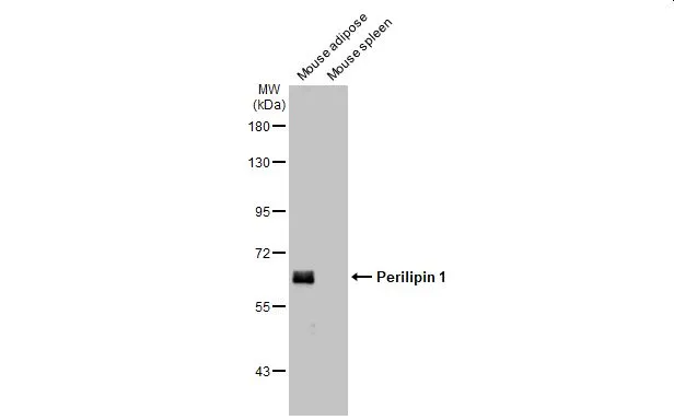
Various tissue extracts (50 μg) were separated by 7.5% SDS-PAGE, and the membrane was blotted with Perilipin 1 antibody (GTX130139) diluted at 1:10000. The HRP-conjugated anti-rabbit IgG antibody (GTX213110-01) was used to detect the primary antibody.

Anti-Perilipin 1 antibody used in Western Blot (WB). GTX130139

GTX130139 WB Image

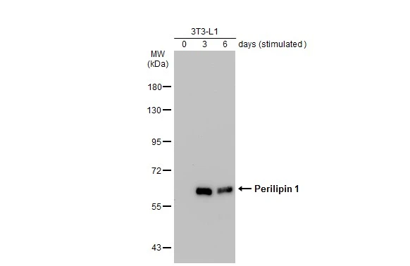
Unstimulatd and stimulatd 3T3-L1 whole cell extracts (20 μg) were separated by 7.5% SDS-PAGE, and the membrane was blotted with Perilipin 1 antibody (GTX130139) diluted at 1:500. The HRP-conjugated anti-rabbit IgG antibody (GTX213110-01) was used to detect the primary antibody. (The differentiation stimulated medium is composed by basal medium, 10% FBS, 50 ug/ml gentamicin, 1 nM L-glutamin, 500 uM IBMX, 1 uM dexamethasone, 2 uM rosiglitazone and 1 ug/ml insulin.)

Anti-Perilipin 1 antibody used in IHC (Paraffin sections) (IHC-P). GTX130139

GTX130139 IHC-P Image

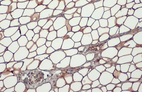
Perilipin 1 antibody detects Perilipin 1 protein at cytoplasm by immunohistochemical analysis.
Sample: Paraffin-embedded mouse white adipocyte.
Perilipin 1 stained by Perilipin 1 antibody (GTX130139) diluted at 1:500.
Antigen Retrieval: Citrate buffer, pH 6.0, 15 min

REFERENCE

REVIEW

There are currently no reviews for Perilipin 1 antibody (GTX130139). Be the first to share your experience with this product.
SDS
PBS.pdf
Glycerol.pdf
Proclin.pdf
Package List Price ($)
$ 399
$ 169