ROBO1 antibody

IHC-Fr analysis of mouse lung tissue using GTX36928 ROBO1 antibody.
Permeabilization : Triton X-100
Dilution : 1:50
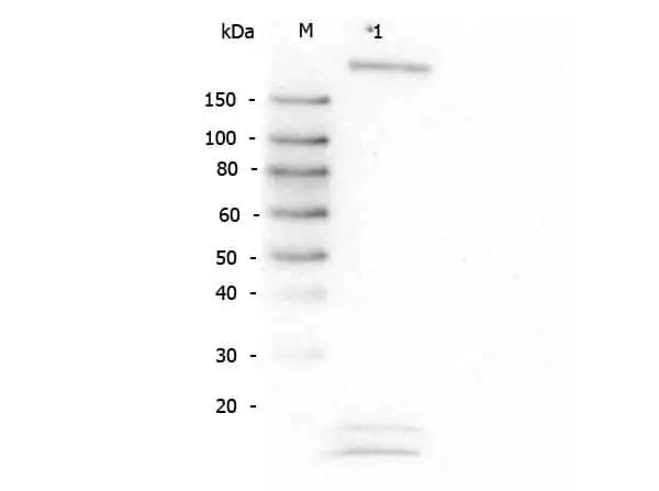
WB analysis of HeLa whole cell lysate using GTX36928 ROBO1 antibody.
Loading : 30 μg
Dilution : 1:1000
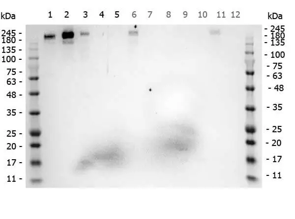
WB analysis of various samples using GTX36928 ROBO1 antibody.
Lane 1 : 293T whole cell lysate
Lane 2 : HeLa whole cell lysate
Lane 3 : MCF-7 whole cell lysate
Lane 4 : Jurkat whole cell lysate
Lane 5 : A431 whole cell lysate
Lane 6 : A549 whole cell lysate
Lane 7 : LNCap whole cell lysate
Lane 8 : Molt-4 whole cell lysate
Lane 9 : Ramos whole cell lysate
Lane 10 : Raji whole cell lysate
Lane 11 : A-172 whole cell lysate
Lane 12 : NIH-3T3 whole cell lysate
Loading : 35 μg
Dilution : 1 μg/mL
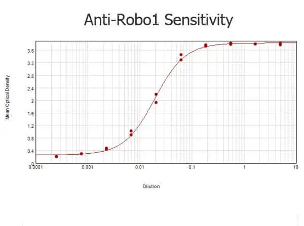
ELISA analysis of BSA-conjugated immunizing peptide using serially diluted GTX36928 ROBO1 antibody.
Coating : 0.1 μg

WB analysis of mouse brain tissue lysate using GTX36928 ROBO1 antibody.
Loading : 35 μg
Dilution : 1:1000
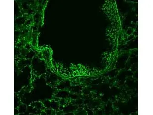
1/50 staining mouse lung tissue sections (adult, frozen 100μm wholemount sections) by IHC-Fr. The tissue was paraformaldehyde fixed and permeabilized with polyethylene glycol tert-octylphenyl ether before incubation with the antibody (GTX36928) for 16 hours at 4ºC.
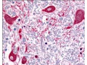
Affinity Purified anti-ROBO1 antibody (GTX36928) was used at a concentration of 5 μg/ml to detect ROBO1 in a variety of tissues including multi-human, multi-brain and multi-cancer slides. This image shows staining of human brain tissue. Tissue was formalin-fixed and paraffin embedded.
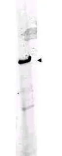
Western blot using Affinity Purified anti-ROBO-1 antibody (GTX36928) shows detection of a band at ~181 kDa corresponding to ROBO-1 present in mouse brain lysate (arrowhead). Approximately 35 μg of lysate was separated by 4-8% SDS-PAGE and transferred onto nitrocellulose. After blocking the membrane was probed with the primary antibody diluted to 1:1,000. Reaction occurred 2h at room temperature followed by washes and reaction with a 1:10,000 dilution of infrared 800 conjugated Gt-a-Rabbit IgG [H&L] MX for 45 min at room temperature. Infrared 800 fluorescence image was captured using the OdysseyR Infrared Imaging System developed by LI-COR.
-
HostRabbit
-
ClonalityPolyclonal
-
IsotypeIgG
-
ApplicationsWB ICC/IF IHC-P IHC-Fr IP ELISA
-
ReactivityHuman, Mouse