RPL32 antibody
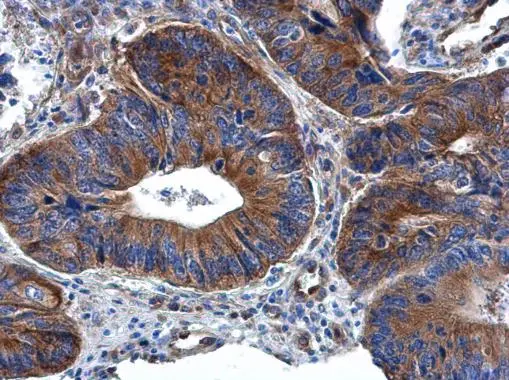
RPL32 antibody detects RPL32 protein at cytoplasm in human colon cancer by immunohistochemical analysis.
Sample: Paraffin-embedded human colon cancer.
RPL32 antibody (GTX130214) diluted at 1:500.
Antigen Retrieval: Citrate buffer, pH 6.0, 15 min
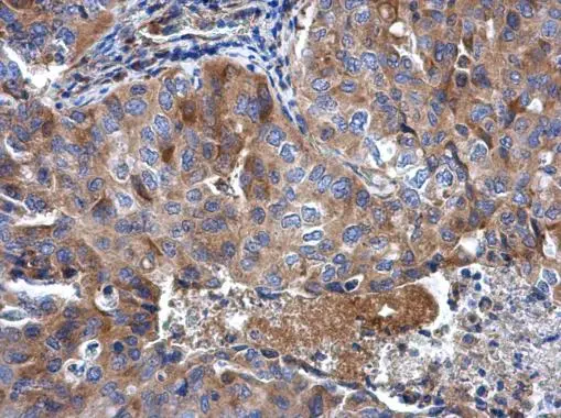
RPL32 antibody detects RPL32 protein at cytoplasm in human lung cancer by immunohistochemical analysis.
Sample: Paraffin-embedded human lung cancer.
RPL32 antibody (GTX130214) diluted at 1:500.
Antigen Retrieval: Citrate buffer, pH 6.0, 15 min
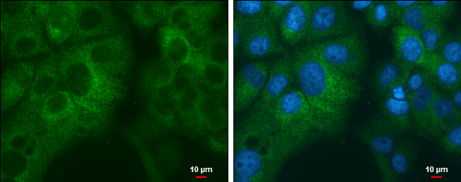
RPL32 antibody detects RPL32 protein at cytoplasm by immunofluorescent analysis.
Sample: MCF7 cells were fixed in 4% paraformaldehyde at RT for 15 min.
Green: RPL32 protein stained by RPL32 antibody (GTX130214) diluted at 1:1000.
Blue: Hoechst 33342 staining.
Scale bar = 10 μm.
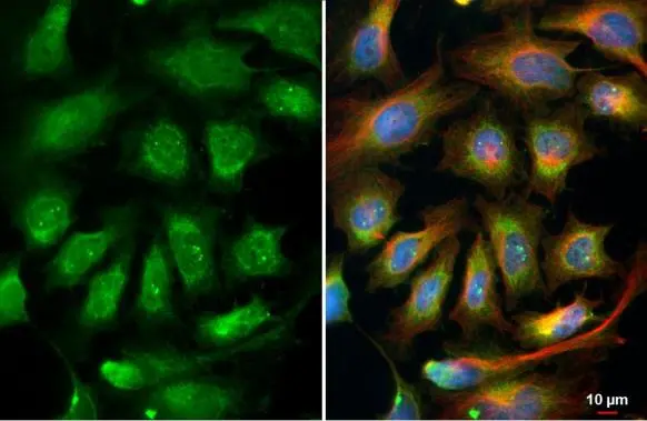
RPL32 antibody detects RPL32 protein at endoplasmic reticulum by immunofluorescent analysis.
Sample: HeLa cells were fixed in 4% paraformaldehyde at RT for 15 min.
Green: RPL32 stained by RPL32 antibody (GTX130214) diluted at 1:500.
Red: alpha Tubulin, a cytoskeleton marker, stained by alpha Tubulin antibody [GT114] (GTX628802) diluted at 1:1000.
Blue: Fluoroshield with DAPI (GTX30920).
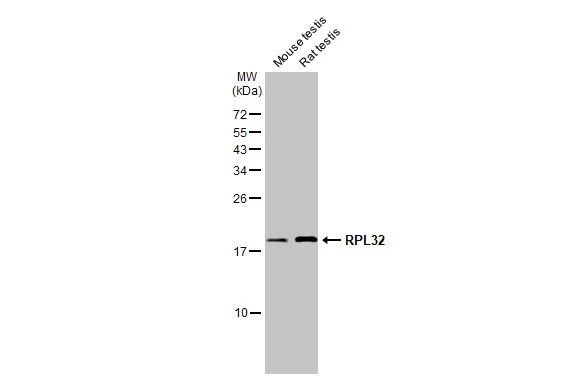
Various tissue extracts (50 μg) were separated by 15% SDS-PAGE, and the membrane was blotted with RPL32 antibody (GTX130214) diluted at 1:1000. The HRP-conjugated anti-rabbit IgG antibody (GTX213110-01) was used to detect the primary antibody.
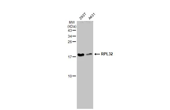
Various whole cell extracts (30 μg) were separated by 15% SDS-PAGE, and the membrane was blotted with RPL32 antibody (GTX130214) diluted at 1:1000. The HRP-conjugated anti-rabbit IgG antibody (GTX213110-01) was used to detect the primary antibody.
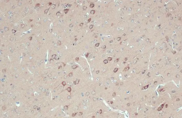
RPL32 antibody detects RPL32 protein at cytoplasm by immunohistochemical analysis.Sample: Paraffin-embedded rat brain.RPL32 stained by RPL32 antibody (GTX130214) diluted at 1:500.Antigen Retrieval: Citrate buffer, pH 6.0, 15 min
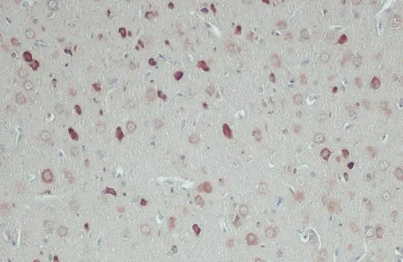
RPL32 antibody detects RPL32 protein at cytoplasm by immunohistochemical analysis.Sample: Paraffin-embedded mouse brain.RPL32 stained by RPL32 antibody (GTX130214) diluted at 1:500.Antigen Retrieval: Citrate buffer, pH 6.0, 15 min
-
HostRabbit
-
ClonalityPolyclonal
-
IsotypeIgG
-
ApplicationsWB ICC/IF IHC-P
-
ReactivityHuman, Mouse, Rat