RPL37 antibody
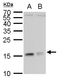
RPL37 antibody detects RPL37 protein by western blot analysis.
A. 30 μg 293T whole cell lysate/extract
B. 30 μg HeLa whole cell lysate/extract
15 % SDS-PAGE
RPL37 antibody (GTX104688) dilution: 1:1000
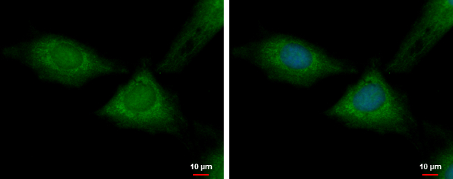
RPL37 antibody detects RPL37 protein at cytoplasm by immunofluorescent analysis.
Sample: HeLa cells were fixed in 4% paraformaldehyde at RT for 15 min.
Green: RPL37 protein stained by RPL37 antibody (GTX104688) diluted at 1:500.
Blue: Hoechst 33342 staining.
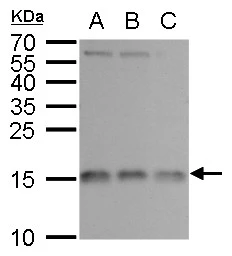
RPL37 antibody detects RPL37 protein by western blot analysis.
A. 30 μg Jurkat whole cell lysate/extract
B. 30 μg Raji whole cell lysate/extract
C. 30 μg NCI-H929 whole cell lysate/extract
15 % SDS-PAGE
RPL37 antibody (GTX104688) dilution: 1:1000
-
HostRabbit
-
ClonalityPolyclonal
-
IsotypeIgG
-
ApplicationsWB ICC/IF
-
ReactivityHuman