RPLP2 antibody

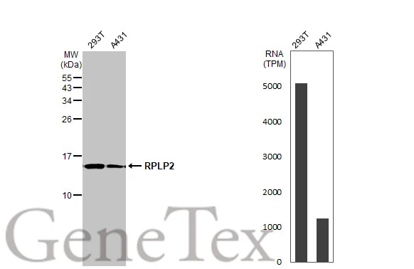
Various whole cell extracts (30 μg) were separated by 15% SDS-PAGE, and the membrane was blotted with RPLP2 antibody (GTX101823) diluted at 1:10000. The HRP-conjugated anti-rabbit IgG antibody (GTX213110-01) was used to detect the primary antibody. Corresponding RNA expression data for the same cell lines are based on Human Protein Atlas program.
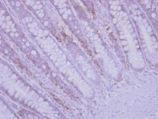
Immunohistochemical analysis of paraffin-embedded human colon carcinoma, using RPLP2(GTX101823) antibody at 1:500 dilution.
Antigen Retrieval: Trilogy™ (EDTA based, pH 8.0) buffer, 15min
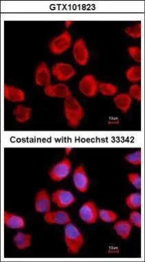
Immunofluorescence analysis of methanol-fixed A431, using RPLP2(GTX101823) antibody at 1:500 dilution.
-
HostRabbit
-
ClonalityPolyclonal
-
IsotypeIgG
-
ApplicationsWB ICC/IF IHC-P
-
ReactivityHuman