Rad21 antibody
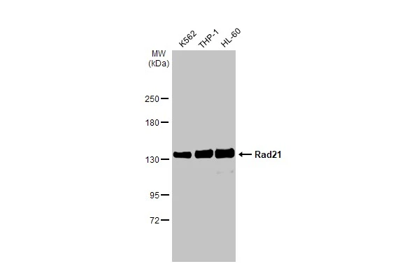
Various whole cell extracts (30 μg) were separated by 5% SDS-PAGE, and the membrane was blotted with Rad21 antibody (GTX106012) diluted at 1:1000. The HRP-conjugated anti-rabbit IgG antibody (GTX213110-01) was used to detect the primary antibody.
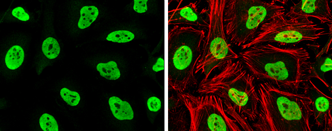
Rad21 antibody detects Rad21 protein at nucleus by immunofluorescent analysis.Sample: HeLa cells were fixed in 4% paraformaldehyde at RT for 15 min.Green: Rad21 stained by Rad21 antibody (GTX106012) diluted at 1:1000.Red: phalloidin, a cytoskeleton marker, diluted at 1:100.
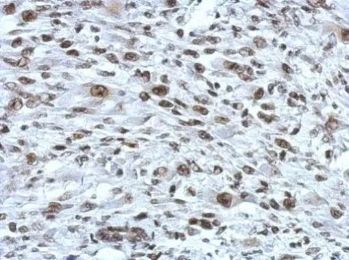
Immunohistochemical analysis of paraffin-embedded HBL435 xenograft, using RAD21(GTX106012) antibody at 1:750 dilution.
Antigen Retrieval: Trilogy™ (EDTA based, pH 8.0) buffer, 15min
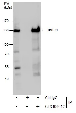
Immunoprecipitation of RAD21 protein from Jurkat whole cell extracts using 5 μg of RAD21 antibody (GTX106012).
Western blot analysis was performed using RAD21 antibody (GTX106012).
EasyBlot anti-Rabbit IgG (GTX221666-01) was used as a secondary reagent.
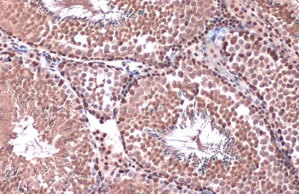
Rad21 antibody detects Rad21 protein at nucleus by immunohistochemical analysis.Sample: Paraffin-embedded mouse testis.Rad21 stained by Rad21 antibody (GTX106012) diluted at 1:500.Antigen Retrieval: Citrate buffer, pH 6.0, 15 min
Immunohistochemical analysis of paraffin-embedded C2C12 xenograft, using RAD21(GTX106012) antibody at 1:750 dilution.
Antigen Retrieval: Trilogy™ (EDTA based, pH 8.0) buffer, 15min
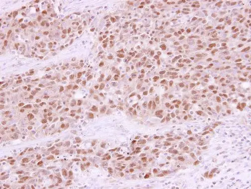
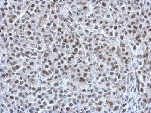
RAD21 antibody detects RAD21 protein at nucleus in human lung papillary adenocarcinoma by immunohistochemical analysis.
Sample: Paraffin-embedded human lung papillary adenocarcinoma.
RAD21 antibody (GTX106012) diluted at 1:250.
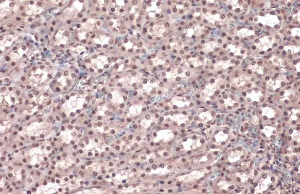
Rad21 antibody detects Rad21 protein at nucleus by immunohistochemical analysis.Sample: Paraffin-embedded rat kidney.Rad21 stained by Rad21 antibody (GTX106012) diluted at 1:500.Antigen Retrieval: Citrate buffer, pH 6.0, 15 min
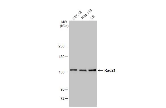
Various whole cell extracts (30 μg) were separated by 5% SDS-PAGE, and the membrane was blotted with Rad21 antibody (GTX106012) diluted at 1:1000. The HRP-conjugated anti-rabbit IgG antibody (GTX213110-01) was used to detect the primary antibody.
-
HostRabbit
-
ClonalityPolyclonal
-
IsotypeIgG
-
ApplicationsWB ICC/IF IHC-P IP ChIP assay
-
ReactivityHuman, Mouse, Rat