Rhodopsin antibody

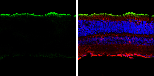
Rhodopsin antibody detects Rhodopsin protein expression by immunohistochemical analysis.
Sample: Frozen sectioned adult mouse retina.
Green: Rhodopsin protein stained by Rhodopsin antibody (GTX129910) diluted at 1:250.
Red: beta Tubulin 3/ TUJ1, stained by beta Tubulin 3/ TUJ1 antibody [GT11710] (GTX631836) diluted at 1:250.
Blue: Fluoroshield with DAPI (GTX30920).
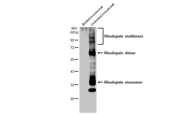
Boiled and unboiled mouse tissue extract (50 μg) were separated by 10% SDS-PAGE, and the membrane was blotted with Rhodopsin antibody (GTX129910) diluted at 1:2000. The HRP-conjugated anti-rabbit IgG antibody (GTX213110-01) was used to detect the primary antibody.
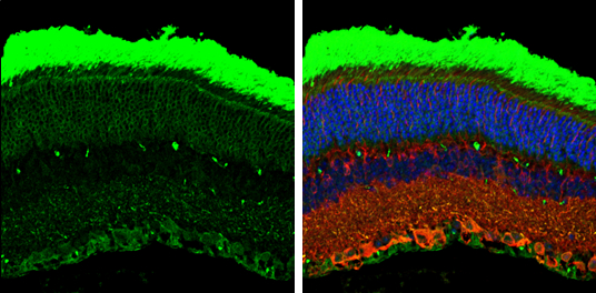
Rhodopsin antibody detects Rhodopsin protein at cell membrane by immunohistochemical analysis.Sample: Paraffin-embedded mouse eye.Green: Rhodopsin stained by Rhodopsin antibody (GTX129910) diluted at 1:250.Red: beta Tubulin 3/ Tuj1 stained by beta Tubulin 3/ Tuj1 antibody [GT1338] (GTX631831) diluted at 1:500.Blue: Fluoroshield with DAPI (GTX30920).
Antigen Retrieval: Citrate buffer, pH 6.0, 15 min
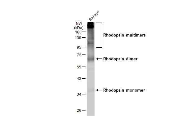
Rat tissue extract (50 μg) was separated by 10% SDS-PAGE, and the membrane was blotted with Rhodopsin antibody (GTX129910) diluted at 1:1000. The HRP-conjugated anti-rabbit IgG antibody (GTX213110-01) was used to detect the primary antibody. Corresponding RNA expression data for the same cell lines are based on Human Protein Atlas program.
-
HostRabbit
-
ClonalityPolyclonal
-
IsotypeIgG
-
ApplicationsWB IHC-P IHC-Fr
-
ReactivityMouse, Rat