S-arrestin antibody
Cat. No. GTX23435
Cat. No. GTX23435
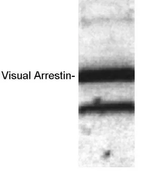
GTX23435 WB Image
WB analysis of recombinant bovine visual arrestin using GTX23435 S-arrestin antibody.
1 / 1
-
HostRabbit
-
ClonalityPolyclonal
-
IsotypeIgG
-
ApplicationsWB ICC/IF IHC
-
ReactivityHuman, Mouse, Sheep, Bovine