S100 beta antibody

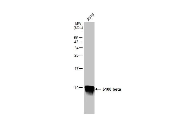
Whole cell extract (30 μg) was separated by 15% SDS-PAGE, and the membrane was blotted with S100 beta antibody (GTX129573) diluted at 1:500. The HRP-conjugated anti-rabbit IgG antibody (GTX213110-01) was used to detect the primary antibody.
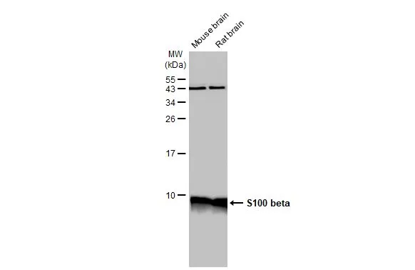
Various tissue extracts (50 μg) were separated by 15% SDS-PAGE, and the membrane was blotted with S100 beta antibody (GTX129573) diluted at 1:1000. The HRP-conjugated anti-rabbit IgG antibody (GTX213110-01) was used to detect the primary antibody.
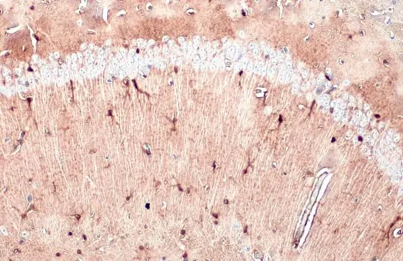
S100 beta antibody detects S100 beta protein at cytoplasm by immunohistochemical analysis.Sample: Paraffin-embedded mouse brain.S100 beta stained by S100 beta antibody (GTX129573) diluted at 1:500.Antigen Retrieval: Citrate buffer, pH 6.0, 15 min
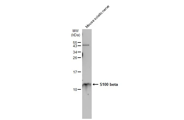
Mouse tissue extract (30 μg) was separated by 15% SDS-PAGE, and the membrane was blotted with S100 beta antibody (GTX129573) diluted at 1:1500. The HRP-conjugated anti-rabbit IgG antibody (GTX213110-01) was used to detect the primary antibody.
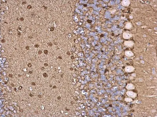
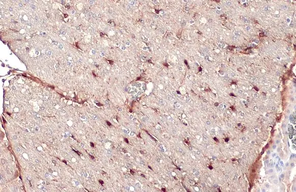
S100B antibody detects S100B protein at glial cell on mouse hind brain by immunohistochemical analysis.
Sample: Paraffin-embedded mouse hind brain.
S100B antibody (GTX129573) dilution: 1:500.
Antigen Retrieval: Trilogy™ (EDTA based, pH 8.0) buffer, 15min
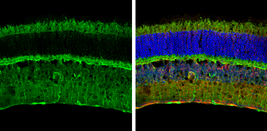
S100 beta antibody detects S100 beta protein expression by immunohistochemical analysis.
Sample: Frozen sectioned adult mouse retina.
Green: S100 beta stained by S100 beta antibody (GTX129573) diluted at 1:250.
Red: beta Tubulin 3/ TUJ1, stained by beta Tubulin 3/ TUJ1 antibody [GT11710] (GTX631836) diluted at 1:250.
Blue: Fluoroshield with DAPI (GTX30920).
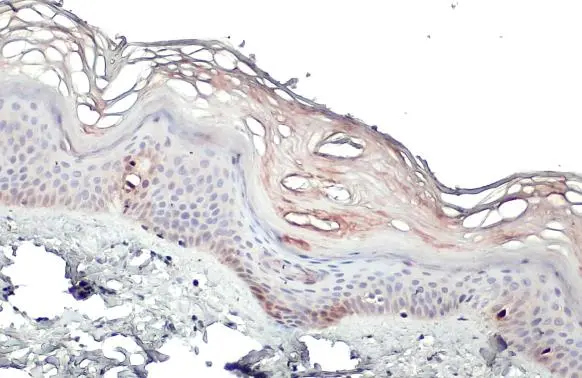
S100 beta antibody detects S100 beta protein at cytoplasm by immunohistochemical analysis.Sample: Paraffin-embedded human skin.S100 beta stained by S100 beta antibody (GTX129573) diluted at 1:500.Antigen Retrieval: Citrate buffer, pH 6.0, 15 min
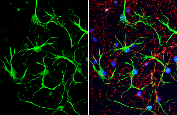
S100 beta antibody detects S100 beta protein at glia cells by immunofluorescent analysis.Sample: DIV10 rat E18 primary cortical neuron and glia cells were fixed in 4% paraformaldehyde at RT for 15 min.Green: S100 beta stained by S100 beta antibody (GTX129573) diluted at 1:500.Red: Tau, stained by Tau antibody [GT287] (GTX634809) diluted at 1:500.Blue: Fluoroshield with DAPI (GTX30920).
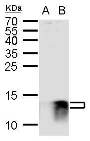
S100B antibody detects S100B protein by western blot analysis.
A. 30 μg 293T whole cell lysate/extract
B. 30 μg whole cell lysate/extract of V5-human S100B-transfected 293T cells
15 % SDS-PAGE
S100B antibody (GTX129573) dilution: 1:1000
S100 beta antibody detects S100 beta protein at cytoplasm by immunohistochemical analysis.Sample: Paraffin-embedded rat brain.S100 beta stained by S100 beta antibody (GTX129573) diluted at 1:500.Antigen Retrieval: Citrate buffer, pH 6.0, 15 min
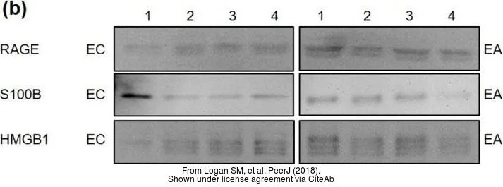
The data was published in the journal PeerJ in 2018. PMID: 29888131
-
HostRabbit
-
ClonalityPolyclonal
-
IsotypeIgG
-
ApplicationsWB ICC/IF IHC-P IHC-Fr
-
ReactivityPolyodon spathula, Human, Mouse, Rat, Squirrel