SA2 antibody

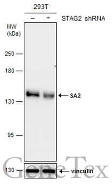
Non-transfected (–) and transfected (+) 293T whole cell extracts (30 μg) were separated by 5% SDS-PAGE, and the membrane was blotted with SA2 antibody (GTX129876) diluted at 1:2000.
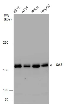
Various whole cell extracts (30 μg) were separated by 5% SDS-PAGE, and the membrane was blotted with SA2 antibody (GTX129876) diluted at 1:1000.
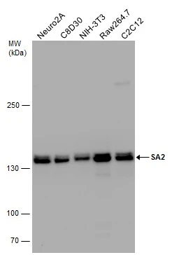
Various whole cell extracts (50 μg) were separated by 5% SDS-PAGE, and the membrane was blotted with SA2 antibody (GTX129876) diluted at 1:1000.
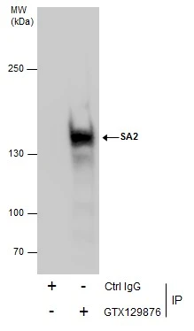
Immunoprecipitation of SA2 protein from 293T whole cell extracts using 5 μg of SA2 antibody (GTX129876).
Western blot analysis was performed using SA2 antibody (GTX129876).
EasyBlot anti-Rabbit IgG (GTX221666-01) was used as a secondary reagent.
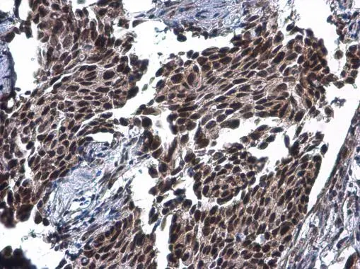
SA2 antibody detects SA2 protein at cytoplasm and nucleus by immunohistochemical analysis.Sample: Paraffin-embedded human breast carcinoma.SA2 stained by SA2 antibody (GTX129876) diluted at 1:500.
Antigen Retrieval: Citrate buffer, pH 6.0, 15 min
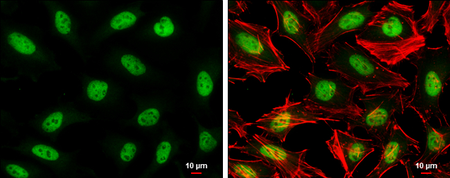
SA2 antibody detects SA2 protein at nucleus by immunofluorescent analysis.Sample: HeLa cells were fixed in 4% paraformaldehyde at RT for 15 min.Green: SA2 stained by SA2 antibody (GTX129876) diluted at 1:500.Red: phalloidin, a cytoskeleton marker, diluted at 1:100.Scale bar= 10μm.
-
HostRabbit
-
ClonalityPolyclonal
-
IsotypeIgG
-
ApplicationsWB ICC/IF IHC-P IP
-
ReactivityHuman, Mouse