APPLICATION
Application Note
*Optimal dilutions/concentrations should be determined by the researcher.
Application |
Recommended Dilution |
1:500-1:3000 |
1:100-1:1000 |
1:100-1:500 |
Not tested in other applications.
Calculated MW
Positive Control
293T , A431 , HeLa , HepG2
Predict Reactivity
Mouse, Rat, Rabbit, Bovine(>80% identity)
PROPERTIES
Form
Liquid
Buffer
PBS, 20% Glycerol
Preservative
0.025% ProClin 300
Storage
Store as concentrated solution. Centrifuge briefly prior to opening vial. For short-term storage (1-2 weeks), store at 4ºC. For long-term storage, aliquot and store at -20ºC or below. Avoid multiple freeze-thaw cycles.
Concentration
1.58 mg/ml (Please refer to the vial label for the specific concentration.)
Antigen Species
Human
Immunogen
Recombinant protein encompassing a sequence within the N-terminus region of human SA2. The exact sequence is proprietary.
Purification
Purified by antigen-affinity chromatography.
Conjugation
Unconjugated
RRID
AB_2886237
Note
For laboratory research use only. Not for any clinical, therapeutic, or diagnostic use in humans or animals. Not for animal or human consumption.
Purchasers shall not, and agree not to enable third parties to, analyze, copy, reverse engineer or otherwise attempt to determine the structure or sequence of the product.
TARGET
Synonyms
stromal antigen 2 , SA-2 , SA2 , SCC3B , bA517O1.1
Cellular Localization
Nucleus
Background
The protein encoded by this gene is a subunit of the cohesin complex, which regulates the separation of sister chromatids during cell division. Targeted inactivation of this gene results in chromatid cohesion defects and aneuploidy, suggesting that genetic disruption of cohesin is a cause of aneuploidy in human cancer. Alternatively spliced transcript variants encoding different isoforms have been found for this gene. [provided by RefSeq, Sep 2013]
Database
Research Area
DATA IMAGES
|
GTX130304 WB Image
Non-transfected (–) and transfected (+) 293T whole cell extracts (30 μg) were separated by 5% SDS-PAGE, and the membrane was blotted with SA2 antibody (GTX130304) diluted at 1:2000.
|
|
GTX130304 IHC-P Image
SA2 antibody detects SA2 protein at nucleus by immunohistochemical analysis.Sample: Paraffin-embedded human breast carcinoma.SA2 stained by SA2 antibody (GTX130304) diluted at 1:500.
Antigen Retrieval: Citrate buffer, pH 6.0, 15 min
|
|
GTX130304 IP Image
Immunoprecipitation of SA2 protein from 293T whole cell extracts using 5 μg of SA2 antibody (GTX130304). Western blot analysis was performed using SA2 antibody (GTX130304) diluted at 1:500. EasyBlot anti-Rabbit IgG (GTX221666-01) was used as a secondary reagent.
|
|
GTX130304 WB Image
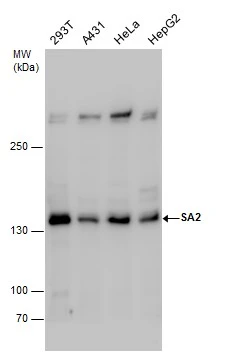
Various whole cell extracts (30 μg) were separated by 5% SDS-PAGE, and the membrane was blotted with SA2 antibody (GTX130304) diluted at 1:1000.
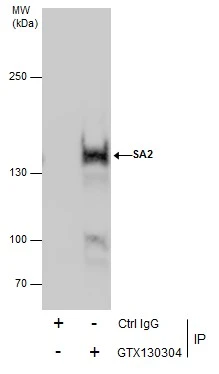
|
REFERENCE
There are currently no references for SA2 antibody (GTX130304). Be the first to share your publications with this product.
REVIEW
There are currently no reviews for SA2 antibody (GTX130304). Be the first to share your experience with this product.