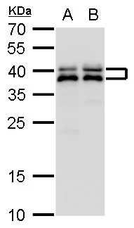
SET antibody

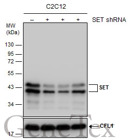
Non-transfected (–) and transfected (+) C2C12 whole cell extracts (30 μg) were separated by 12% SDS-PAGE, and the membrane was blotted with SET antibody (GTX113834) diluted at 1:1000.
Observed SET protein is larger than the predicted M.W., possibly due to post-translational modifications.
Reference: Exp Cell Res. 1998 May 1;240(2):274-81.
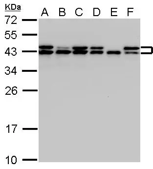
SET antibody detects SET protein by Western blot analysis.
A. 30 μg A431 whole cell lysate/extract whole cell lysate/extract
B. 30 μg H1299 whole cell lysate/extract
C. 30 μg HeLa whole cell lysate/extract
D. 30 μg HepG2 whole cell lysate/extract
E. 30 μg Molt-4 whole cell lysate/extract
F. 30 μg Raji whole cell lysate/extract
12 % SDS-PAGE
SET antibody (GTX113834) dilution: 1:1000 SET antibody detects SET protein by Western blot analysis.
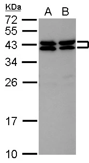
SET antibody detects SET protein by Western blot analysis.
A. 30 μg JC whole cell lysate/extract
B. 30 μg BCL-1 whole cell lysate/extract
12 % SDS-PAGE
SET antibody (GTX113834) dilution: 1:3000
SET antibody detects SET protein by Western blot analysis.
A. 30 μg Jurkat whole cell lysate/extract
B. 30 μg Raji whole cell lysate/extract
C. 30 μg K562 whole cell lysate/extract
D. 30 μg THP-1 whole cell lysate/extract
E. 30 μg HL-60 whole cell lysate/extract
12 % SDS-PAGE
SET antibody (GTX113834) dilution: 1:10000
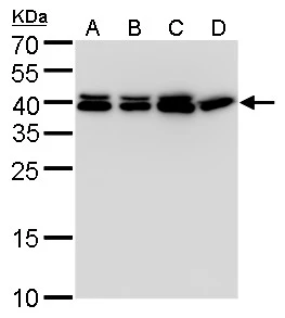
SET antibody detects SET protein by Western blot analysis.
A. 30 μg 293T whole cell lysate/extract
B. 30 μg A431 whole cell lysate/extract
C. 30 μg HeLa whole cell lysate/extract
D. 30 μg HepG2 whole cell lysate/extract
12 % SDS-PAGE
SET antibody (GTX113834) dilution: 1:2500
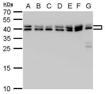
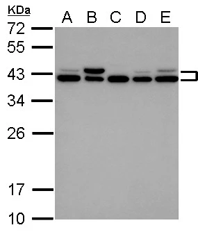
SET antibody detects SET protein by western blot analysis.
A. 30 μg Neuro2A whole cell lysate/extract
B. 30 μg GL261 whole cell lysate/extract
C. 30 μg C8D30 whole cell lysate/extract
D. 30 μg NIH-3T3 whole cell lysate/extract
E. 30 μg BCL-1 whole cell lysate/extract
F. 30 μg Raw 264.7 whole cell lysate/extract
G. 30 μg C2Cl2 whole cell lysate/extract
12 % SDS-PAGE
SET antibody (GTX113834) dilution: 1:1000
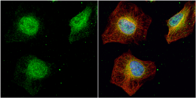
SET antibody detects SET protein at cytoplasm and nucleus by immunofluorescent analysis.
Sample: HeLa cells were fixed in 4% paraformaldehyde at RT for 15 min.
Green: SET protein stained by SET antibody (GTX113834) diluted at 1:500.
Red: alpha Tubulin, a cytoskeleton marker, stained by alpha Tubulin antibody [GT114] (GTX628802) diluted at 1:1000.
Blue: Hoechst 33342 staining.
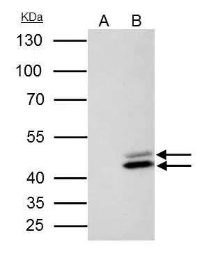
SET antibody immunoprecipitates SET protein in IP experiments.
IP samples: HeLa whole cell extract
A. Control with 4 μg of preimmune Rabbit IgG
B. Immunoprecipitation of SET protein by 4 μg SET antibody (GTX113834)
10 % SDS-PAGE
The immunoprecipitated SET protein was detected by SET antibody (GTX113834) diluted at 1:500.
[EasyBlot anti-rabbit IgG (GTX221666-01) was used as a secondary reagent]
SET antibody detects SET protein by western blot analysis.
A. 30 μg PC-12 whole cell lysate/extract
B. 30 μg Rat2 whole cell lysate/extract
12 % SDS-PAGE
SET antibody (GTX113834) dilution: 1:1000
-
HostRabbit
-
ClonalityPolyclonal
-
IsotypeIgG
-
ApplicationsWB ICC/IF IP
-
ReactivityHuman, Mouse, Rat