SLIT2 antibody


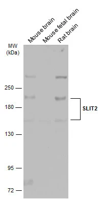
Various tissue extracts (50 μg) were separated by 5% SDS-PAGE, and the membrane was blotted with SLIT2 antibody (GTX118220) diluted at 1:1000. The HRP-conjugated anti-rabbit IgG antibody (GTX213110-01) was used to detect the primary antibody.
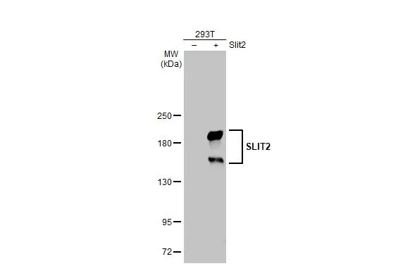
Non-transfected (–) and transfected (+) 293T whole cell extracts (30 μg) were separated by 5% SDS-PAGE, and the membrane was blotted with SLIT2 antibody (GTX118220) diluted at 1:2000. The HRP-conjugated anti-rabbit IgG antibody (GTX213110-01) was used to detect the primary antibody.
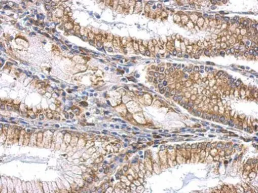
SLIT2 antibody detects SLIT2 protein at cytosol and membrane on gastric carcinoma by immunohistochemical analysis.
Sample: Paraffin-embedded human gastric carcinoma.
SLIT2 antibody (GTX118220) dilution: 1:500.
Antigen Retrieval: Trilogy™ (EDTA based, pH 8.0) buffer, 15min
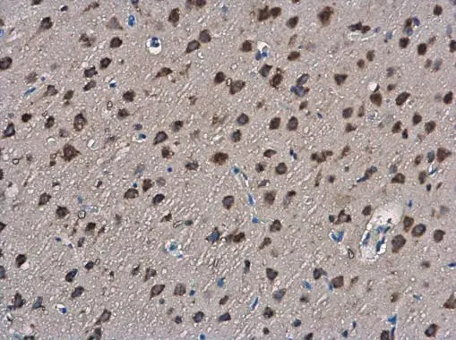
SLIT2 antibody detects SLIT2 protein at cytoplasm in rat brain by immunohistochemical analysis.
Sample: Paraffin-embedded rat brain.
SLIT2 antibody (GTX118220) diluted at 1:400.
Antigen Retrieval: Citrate buffer, pH 6.0, 15 min
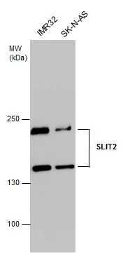
SLIT2 antibody detects SLIT2 protein by western blot analysis. Various whole cell extracts (30 μg) were separated by 5% SDS-PAGE, and the membrane was blotted with SLIT2 antibody (GTX118220) diluted at 1:1000. The HRP-conjugated anti-rabbit IgG antibody (GTX213110-01) was used to detect the primary antibody.
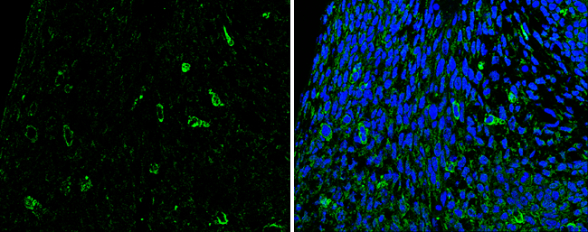
SLIT2 antibody detects SLIT2 protein at cytoplasm in mouse fetal brain by immunohistochemical analysis.
Sample: Paraffin-embedded mouse fetal brain.
Green: SLIT2 antibody (GTX118220) diluted at 1:200. The signal was developed using goat anti-rabbit IgG antibody (Dylight488) (GTX213110-04).
Blue: Nuclear staining with Hoechst 33342.
Antigen Retrieval: Citrate buffer, pH 6.0, 15 min
-
HostRabbit
-
ClonalityPolyclonal
-
IsotypeIgG
-
ApplicationsWB IHC-P
-
ReactivityHuman, Mouse, Rat