APPLICATION
Application Note
*Optimal dilutions/concentrations should be determined by the researcher.
Application |
Dilution |
1:500-1:3000 |
1:100-1:1500 |
Assay dependent |
Not tested in other applications.
Calculated MW
Positive Control
treated MCF-7 whole cell extract
Predict Reactivity
Pig, Xenopus laevis(>80% identity)
PROPERTIES
Form
Liquid
Buffer
1XPBS, 1% BSA, 20% Glycerol (pH7). 0.025% ProClin 300 was added as a preservative.
Storage
Store as concentrated solution. Centrifuge briefly prior to opening vial. For short-term storage (1-2 weeks), store at 4ºC. For long-term storage, aliquot and store at -20ºC or below. Avoid multiple freeze-thaw cycles.
Concentration
0.44 mg/ml (Please refer to the vial label for the specific concentration.)
Antigen Species
Human
Immunogen
Carrier-protein conjugated synthetic peptide corresponding to residues around human SMAD2 (phospho Ser465/Ser467). The exact sequence is proprietary.
Purification
Purified by antigen-affinity chromatography.
Conjugation
Unconjugated
RRID
AB_2887051
Note
For laboratory use only. Not for any clinical, therapeutic, or diagnostic use in humans or animals. Not for animal or human consumption.
TARGET
Synonyms
SMAD family member 2 , JV18 , JV18-1 , MADH2 , MADR2 , hMAD-2 , hSMAD2
Cellular Localization
Cytoplasm , Nucleus
Background
The protein encoded by this gene belongs to the SMAD, a family of proteins similar to the gene products of the Drosophila gene 'mothers against decapentaplegic' (Mad) and the C. elegans gene Sma. SMAD proteins are signal transducers and transcriptional modulators that mediate multiple signaling pathways. This protein mediates the signal of the transforming growth factor (TGF)-beta, and thus regulates multiple cellular processes, such as cell proliferation, apoptosis, and differentiation. This protein is recruited to the TGF-beta receptors through its interaction with the SMAD anchor for receptor activation (SARA) protein. In response to TGF-beta signal, this protein is phosphorylated by the TGF-beta receptors. The phosphorylation induces the dissociation of this protein with SARA and the association with the family member SMAD4. The association with SMAD4 is important for the translocation of this protein into the nucleus, where it binds to target promoters and forms a transcription repressor complex with other cofactors. This protein can also be phosphorylated by activin type 1 receptor kinase, and mediates the signal from the activin. Alternatively spliced transcript variants encoding the same protein have been observed. [provided by RefSeq]
Database
Research Area
DATA IMAGES
|
GTX133614 ICC/IF Image
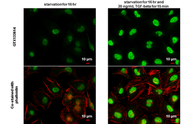
SMAD2 (phospho Ser465/Ser467) antibody detects SMAD2 (phospho Ser465/Ser467) protein at nucleus by immunofluorescent analysis.Sample: Mock and treated HeLa cells were fixed in 4% paraformaldehyde at RT for 15 min.Green: SMAD2 (phospho Ser465/Ser467) stained by SMAD2 (phospho Ser465/Ser467) antibody (GTX133614) diluted at 1:1500.Red: phalloidin, a cytoskeleton marker, diluted at 1:100.Scale bar= 10 μm.
|
|
GTX133614 WB Image
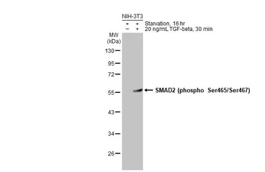
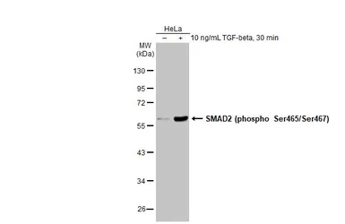
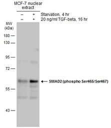
Untreated (–) and treated (+) MCF-7 whole cell extract (30 μg) were separated by 10% SDS-PAGE, and the membrane was blotted with SMAD2 (phospho Ser465/Ser467) antibody (GTX133614) diluted at 1:1000. The HRP-conjugated anti-rabbit IgG antibody (GTX213110-01) was used to detect the primary antibody.
|
REFERENCE
REVIEW
There are currently no reviews for SMAD2 (phospho Ser465/Ser467) antibody (GTX133614). Be the first to share your experience with this product.