SP1 antibody
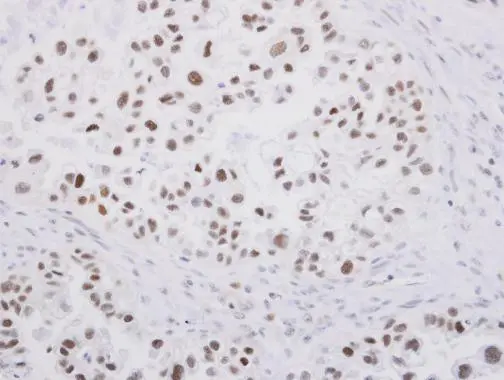
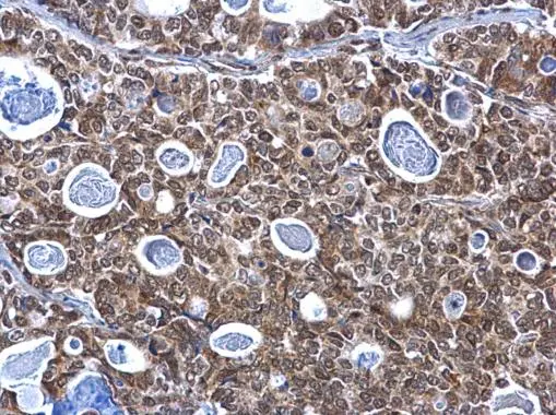
SP1 antibody detects SP1 protein at nucleus on human ovarian carcinoma by immunohistochemical analysis.
Sample: Paraffin-embedded human ovarian carcinoma.
SP1 antibody (GTX103031) diluted at 1:250.
Antigen Retrieval: Trilogy™ (EDTA based, pH 8.0) buffer, 15min
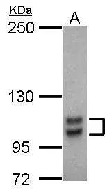
Sample (30 ug of whole cell lysate)
A: THP-1
5% SDS PAGE
GTX103031 diluted at 1:1000
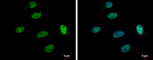
SP1 antibody detects SP1 protein at nucleus by immunofluorescent analysis.
Sample: A549 cells were fixed in 4% paraformaldehyde at RT for 15 min.
Green: SP1 protein stained by SP1 antibody (GTX103031) diluted at 1:500.
Blue: Hoechst 33342 staining.
Scale bar = 6 μm.
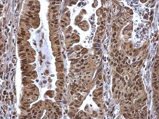
SP1 antibody detects SP1 protein at nucleus on human gastric carcinoma by immunohistochemical analysis.
Sample: Paraffin-embedded human gastric carcinoma.
SP1 antibody (GTX103031) dilution: 1:500.
Antigen Retrieval: Trilogy™ (EDTA based, pH 8.0) buffer, 15min
SP1 antibody detects SP1 protein at nucleus on human gastric carcinoma by immunohistochemical analysis.
Sample: Paraffin-embedded human gastric carcinoma.
SP1 antibody (GTX103031) dilution: 1:500.
Antigen Retrieval: Trilogy™ (EDTA based, pH 8.0) buffer, 15min
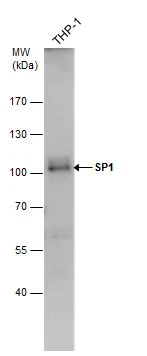
SP1 antibody detects SP1 protein by western blot analysis. Whole cell extracts (30 μg) was separated by 7.5% SDS-PAGE, and the membrane was blotted with SP1 antibody (GTX103031) diluted by 1:500.
-
HostRabbit
-
ClonalityPolyclonal
-
IsotypeIgG
-
ApplicationsWB ICC/IF IHC-P
-
ReactivityHuman