SP3 antibody


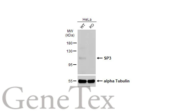
Wild-type (WT) and SP3 knockout (KO) HeLa cell extracts (30 μg) were separated by 7.5% SDS-PAGE, and the membrane was blotted with SP3 antibody (GTX129426) diluted at 1:1000. The HRP-conjugated anti-rabbit IgG antibody (GTX213110-01) was used to detect the primary antibody. Corresponding RNA expression data for the same cell lines are based on Human Protein Atlas program.
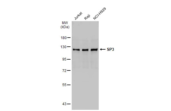
Various whole cell extracts (30 μg) were separated by 7.5% SDS-PAGE, and the membrane was blotted with SP3 antibody (GTX129426) diluted at 1:1000. The HRP-conjugated anti-rabbit IgG antibody (GTX213110-01) was used to detect the primary antibody.
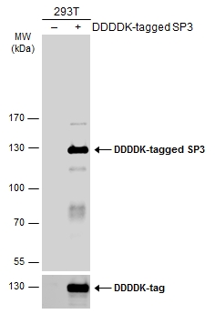
Non-transfected (–) and transfected (+) 293T whole cell extracts (30 μg) were separated by 7.5% SDS-PAGE, and the membrane was blotted with SP3 antibody (GTX129426) diluted at 1:1000. The HRP-conjugated anti-rabbit IgG antibody (GTX213110-01) was used to detect the primary antibody.
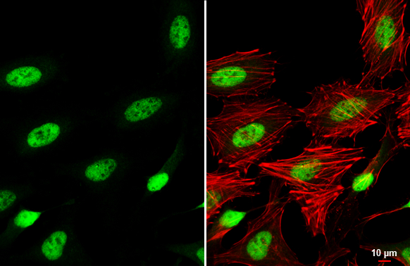
SP3 antibody detects SP3 protein at nucleus by immunofluorescent analysis.Sample: HeLa cells were fixed in 4% paraformaldehyde at RT for 15 min.Green: SP3 stained by SP3 antibody (GTX129426) diluted at 1:500.Red: phalloidin, a cytoskeleton marker, diluted at 1:200.Scale bar= 10 μm.
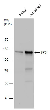
SP3 antibody detects SP3 protein by western blot analysis.
A. 30 μg Jurkat whole cell extract
B. 30 μg Jurkat nuclear extract
7.5 % SDS-PAGE
SP3 antibody (GTX129426) dilution: 1:1000
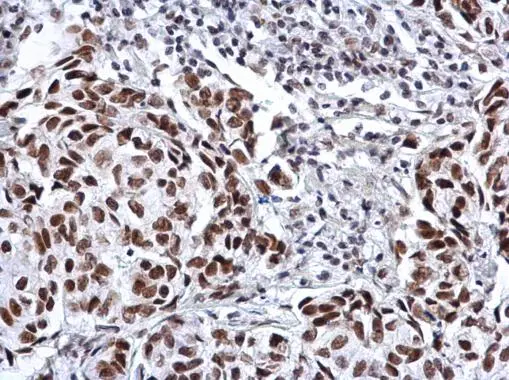
SP3 antibody detects SP3 protein at nucleus by immunohistochemical analysis.Sample: Paraffin-embedded human lung cancer.SP3 stained by SP3 antibody (GTX129426) diluted at 1:500.Antigen Retrieval: Citrate buffer, pH 6.0, 15 min
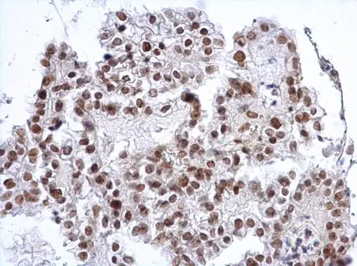
SP3 antibody detects SP3 protein at nucleus on human breast carcinoma by immunohistochemical analysis.
Sample: Paraffin-embedded human breast carcinoma.
SP3 antibody (GTX129426) dilution: 1:1000.
Antigen Retrieval: Trilogy™ (EDTA based, pH 8.0) buffer, 15min
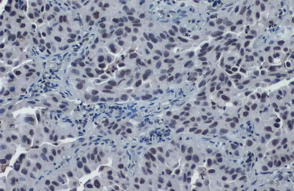
SP3 antibody detects SP3 protein at nucleus on human ovarian carcinoma by immunohistochemical analysis.
Sample: Paraffin-embedded human ovarian carcinoma.
SP3 antibody (GTX129426) dilution: 1:1000.
Antigen Retrieval: Trilogy™ (EDTA based, pH 8.0) buffer, 15min
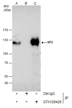
Immunoprecipitation of SP3 protein from Jurkat nuclear extracts using 5 μg of SP3 antibody (GTX129426).
Western blot analysis was performed using SP3 antibody (GTX129426) diluted at 1:500.
EasyBlot anti-Rabbit IgG (GTX221666-01) was used as a secondary reagent.
-
HostRabbit
-
ClonalityPolyclonal
-
IsotypeIgG
-
ApplicationsWB ICC/IF IHC-P IP
-
ReactivityHuman