SPANXC antibody
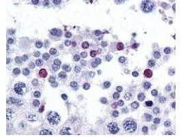
GeneTex's affinity purified anti-SPANX-C antibody was used at 2.5 μg/ml to detect signal in a variety of tissues including multi-human, multi-brain and multi-cancer slides. This image shows moderate positive staining of human sperm and spermatids at 60X. Tissue was formalin-fixed and paraffin embedded. The image shows localization of the antibody as the precipitated red signal, with a hematoxylin purple nuclear counterstain.
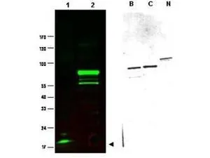
Western blot using GeneTex's Protein A purified anti-SPANX-C antibody shows detection of a band at ~17 kDa corresponding to SPANX-C present in a nuclear extract from VWM105 cells (left panel, arrowhead). VWM105 cells are derived from a human melanoma and are positive for SPANX proteins. Lane 2 shows reactivity with a purified recombinant SPANX-C fusion protein. The right panel shows similar reactivity with purified recombinant SPANX-B, SPANX-C and SPANX-N proteins. Proteins were separated by SDS-PAGE, transferred to nitrocellulose, and probed with the primary antibody diluted to 1:1,000. IRDye™800 conjugated goat anti-Rabbit IgG [H&L] was used (left). IRDye is a trademark of LI-COR, Inc. Size estimation was made by comparison to prestained MW markers as indicated.
-
HostRabbit
-
ClonalityPolyclonal
-
IsotypeIgG
-
ApplicationsWB IHC-P ELISA
-
ReactivityHuman