SR-BI antibody
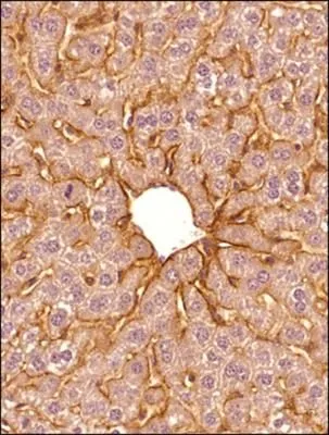
IHC-P analysis of mouse liver tissue using GTX20396 SR-BI antibody.
Dilution : 1:300
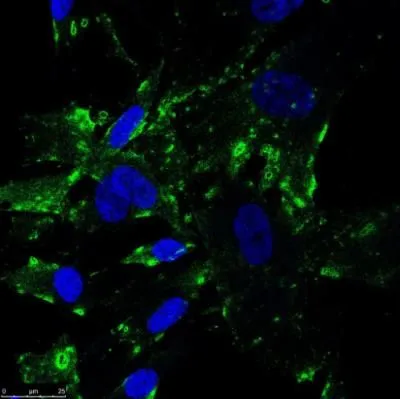
ICC/IF analysis of human fibroblasts using GTX20396 SR-BI antibody.
Green : primary antibody
Blue : DAPI
Dilution : 1:100
Fixation : 4% PFA
Permeabilization : 0.2% Tween
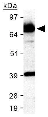
WB analysis of mouse liver tissue lysate using GTX20396 SR-BI antibody.
Loading : 25μg
Dilution : 1:1000
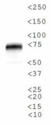
WB analysis of human adrenal tissue using GTX20396 SR-BI antibody.
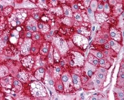
IHC-P analysis of human adrenal cortex tissue using GTX20396 SR-BI antibody.

WB analysis of HeLa cell lysate using GTX20396 SR-BI antibody.

IHC-P analysis of mouse liver tissue using GTX20396 SR-BI antibody.
Dilution : 1:300
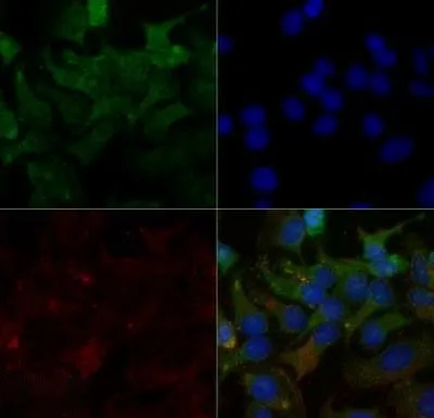
ICC/IF analysis of HeLa cells using GTX20396 SR-BI antibody.
Green : primary antibody
Red : Tubulin
Blue : DAPI
Dilution : 2 μg/ml
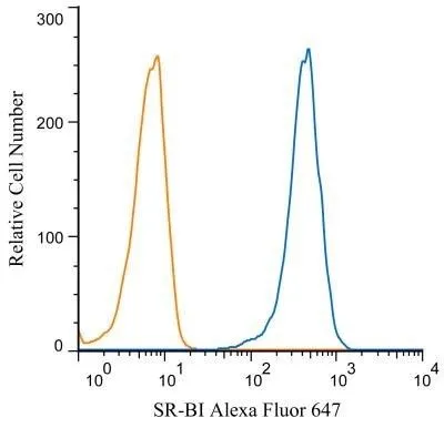
FACS (Intracellular staining) analysis of HeLa cells using GTX20396 SR-BI antibody.
Blue : Primary antibody
Orange : isotype control
Dilution : 2 μg/mL
Fixation : 4% PFA
Permibilization : 0.1% saponin
-
HostRabbit
-
ClonalityPolyclonal
-
IsotypeIgG
-
ApplicationsWB ICC/IF IHC-P IHC-Fr FCM IP
-
ReactivityHuman, Mouse, Rat, Rabbit, Hamster, Golden Syrian Hamster, Mustelid, Primate