STK3 antibody

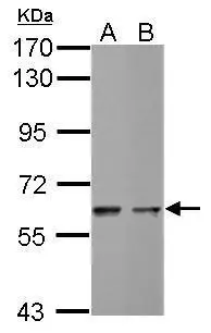
Sample (30 ug of whole cell lysate)
A: THP-1
B: NCI-H929
7.5% SDS PAGE
GTX111494 diluted at 1:1000
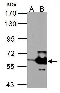
Sample (30 ug of whole cell lysate)
A: Non-transfected 293T lysates
B: STK3 transfected 293T lysates
7.5% SDS PAGE
GTX111494 diluted at 1:5000
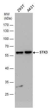
STK3 antibody detects STK3 protein by western blot analysis. Various whole cell extracts (30 μg) were separated by 10% SDS-PAGE, and the membrane was blotted with STK3 antibody (GTX111494) diluted by 1:1000.
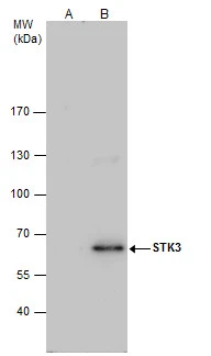
STK3 antibody immunoprecipitates STK3 protein in IP experiments.
IP samples: HeLa whole cell extract
A. Control with 4 μg of preimmune Rabbit IgG
B. Immunoprecipitation of STK3 protein by 4 μg STK3 antibody (GTX111494)
7.5 % SDS-PAGE
The immunoprecipitated STK3 protein was detected by STK3 antibody (GTX111494) diluted at 1:500.
[EasyBlot anti-rabbit IgG (GTX221666-01) was used as a secondary reagent]
-
HostRabbit
-
ClonalityPolyclonal
-
IsotypeIgG
-
ApplicationsWB IP
-
ReactivityHuman