Semaphorin 3A antibody
Cat. No. GTX130671
Cat. No. GTX130671
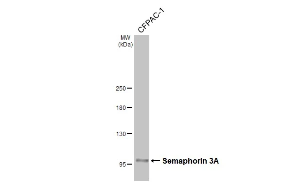
GTX130671 WB Image
Whole cell extract (30 μg) was separated by 5% SDS-PAGE, and the membrane was blotted with Semaphorin 3A antibody (GTX130671) diluted at 1:1000. The HRP-conjugated anti-rabbit IgG antibody (GTX213110-01) was used to detect the primary antibody.
1 / 1
-
HostRabbit
-
ClonalityPolyclonal
-
IsotypeIgG
-
ApplicationsWB
-
ReactivityHuman