TIGAR antibody
Cat. No. GTX31307
Cat. No. GTX31307
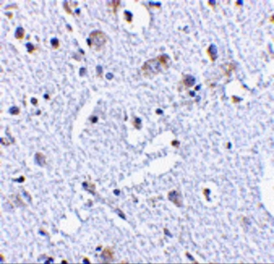
GTX31307 IHC-P Image
IHC-P analysis of human brain tissue using GTX31307 TIGAR antibody.
Working concentration : 20 μg/ml
1 / 3
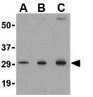
GTX31307 WB Image
WB analysis of MCF7 cell lysate using GTX31307 TIGAR antibody.
Working concentration : (A) 0.5, (B) 1, and (C) 2 μg/ml
2 / 3
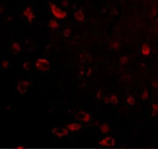
GTX31307 IHC-P Image
IHC-P analysis of human brain tissue using GTX31307 TIGAR antibody.
Working concentration : 2.5 μg/ml
3 / 3
-
HostRabbit
-
ClonalityPolyclonal
-
IsotypeIgG
-
ApplicationsWB IHC-P ELISA
-
ReactivityHuman, Mouse