TMS1 antibody
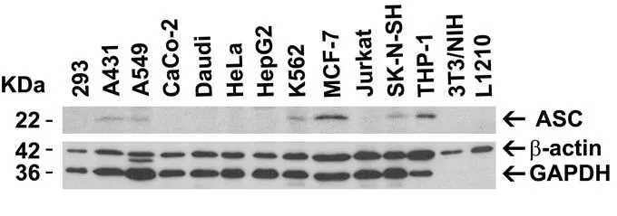
WB analysis of various samples using GTX28394 TMS1 antibody.
Dilution : 2μg/mL
Loading : 15μg of lysates per lane
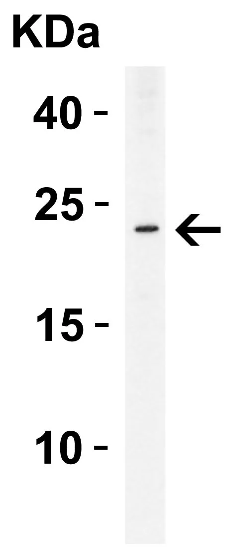
WB analysis of THP-1 cell lysate using GTX28394 TMS1 antibody.
Dilution : 2μg/mL
Loading : 15μg of lysates per lane
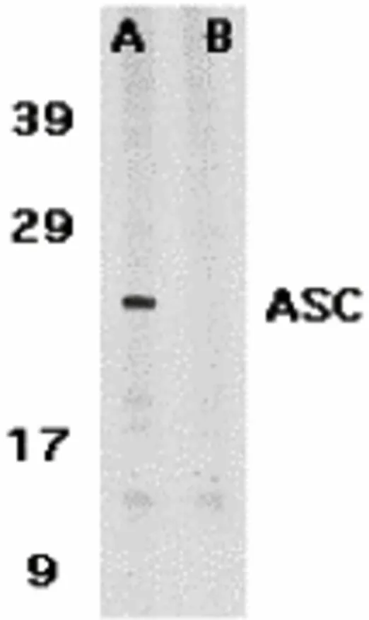
WB analysis of HL-60 cell lysate using GTX28394 TMS1 antibody in the absence (A) or presence (B) of blocking peptide.
Dilution : 1μg/mL
Loading : 15μg of lysates per lane
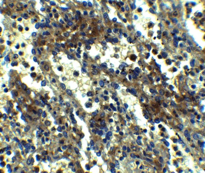
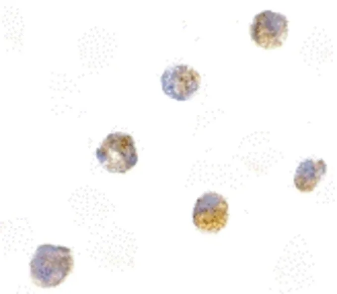
IHC-P analysis of formaldehyde fixed Human Spleen Tissue using GTX28394 TMS1 antibody.
Antigen retreival : Heat mediation with a citrate buffer (pH6)
Dilution : 2.5μg/mL
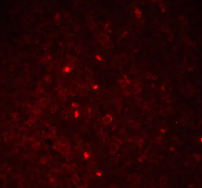
ICC/IF analysis of formaldehyde-fixed HL-60 cells using GTX28394 TMS1 antibody.
Antigen retrieval : Heat mediation with a citrate buffer (pH6)
Dilution : 5μg/mL
IHC-P analysis of 4% PFA-fixed Human Spleen Tissue using GTX28394 TMS1 antibody.
Dilution : 20μg/mL
-
HostRabbit
-
ClonalityPolyclonal
-
IsotypeIgG
-
ApplicationsWB ICC/IF IHC-P ELISA
-
ReactivityHuman